Old Browser
This page has been recently translated and is available in French now.
Looks like you're visiting us from {countryName}.
Would you like to stay on the current country site or be switched to your country?



Flow cytometric analysis of CD95 expression on human peripheral blood lymphocytes. Whole blood was stained with either Purified NA/LE Mouse Anti-Human CD95 (Cat. No. 555670; solid line histogram) or Purified NA/LE Mouse IgG1 κ Isotype Control (Cat. No. 554721; dashed line histogram), followed by FITC Goat Anti-Mouse IgG/IgM (Cat. No. 555988). Erythrocytes were lysed with BD Pharm Lyse™ Lysing Buffer (Cat. No. 555899).


BD Pharmingen™ Purified NA/LE Mouse Anti-Human CD95

Regulatory Status Legend
Any use of products other than the permitted use without the express written authorization of Becton, Dickinson and Company is strictly prohibited.
Preparation And Storage
Recommended Assay Procedures
Investigators are advised that the following procedure is not routinely tested for this material.
Induction of Apoptosis Using Purified Mouse Anti-Human CD95 (Clone DX2) [MN 555670]
Additional Materials Needed:
Positive control cell line (e.g., Daudi, HPB-ALL, Jurkat)
Recombinant Protein G (rProt G) (SIGMA, Cat. No. P4689)
96-well microtiter plate
IMDM or RPMI 1640 medium with 10% heat-inactivated fetal bovine serum (FBS), 1% L-glutamine, 1% antibiotics (FBS medium)
Procedure:
1. Maintain cells in culture with 10% FBS medium; change the medium one day before starting the induction of apoptosis. Harvest and pellet the cells, resuspend in 10% FBS medium at a density of 0.5-1.0 x 10^6 cells/ml.
2. In a 96-well microtiter plate, add 2.5 µg/50 µl CD95 NA/LE™, 0.5 µg/50 µl rProt G, 0.5-1.0 x 10^5 cells and 10% FBS medium to a total volume of 200 µl. Negative controls should consist of: 1) 0.5-1.0 x 10^5 cells with 10% FBS medium alone (200 µl total volume), and 2) 0.5-1.0 x 10^5 cells with 0.5 µg/50 µl rProtG and 10% FBS medium.
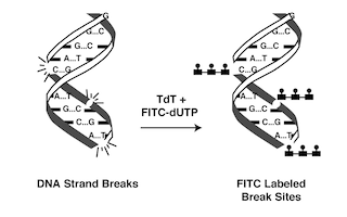
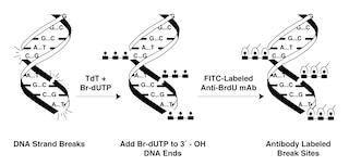
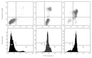
3. Incubate the 96-well plate at 37°C, 5% CO2, for 12-24 hours. Traditionally, apoptosis has been observed by light microscopy, MTT, or gel electrophoresis (DNA fragmentation). Investigators may be interested in several products offered by BD Biosciences that may be used for the detection of apoptotic cells by flow cytometry: ApoDirect™ Kit (Cat. No. 556381), ApoBRDU™ Kit (Cat. No. 556405) and Annexin V-FITC (Cat. No. 556420 or 556419). Because these methods vary in sensitivity, it may be necessary to titer the rProtG and/or CD95 NA/LE™ to obtain optimal results.
Notes:
The suggestions given in this procedure are based on conditions that were found to be optimal for the induction of apoptosis by anti-human CD95 (Fas). Studies have shown that the addition of protein G can significantly enhance the efficiency of DX2 in this type of functional assay. It is important to note that there is a great deal of variation between cell lines in the level of apoptosis that can be induced through the Fas receptor. It has been reported that, although some cells express the Fas antigen, they do not necessarily undergo Fas-mediated apoptosis (i.e., the Fas antigen expressed may be anti-Fas sensitive or insensitive). Daudi, Jurkat, and HPB-ALL cells are good positive controls as they are strongly induced by anti-Fas to undergo apoptosis.
Product Notices
- Since applications vary, each investigator should titrate the reagent to obtain optimal results.
- An isotype control should be used at the same concentration as the antibody of interest.
- Species cross-reactivity detected in product development may not have been confirmed on every format and/or application.
- Please refer to www.bdbiosciences.com/us/s/resources for technical protocols.
Companion Products
.png?imwidth=320)
The DX2 monoclonal antibody specifically binds to the human Fas antigen (also called APO-1). This 45 kDa type I transmembrane glycoprotein was designated as CD95 at the Fifth HLDA Workshop. Fas is a member of the TNF-receptor superfamily and is also known as Tumor necrosis factor receptor superfamily member 6 (TNFRSF6). It is differentially expressed on a variety of normal and neoplastic cells. These include some undifferentiated thymocytes, and activated T and B lymphocytes, natural killer (NK) cells, monocytes, neutrophils, fibroblasts, and cell lines. CD95 is preferentially expressed on CD45RO-positive memory T lymphocytes and γ/δ T lymphocytes. The Fas/CD95 antigen is a polypeptide that plays a role in the programmed sequence of events leading to cell death, termed apoptosis. Crosslinking CD95 with DX2 antibody delivers an apoptotic signal indicating that DX2 recognizes a functional epitope of the CD95 antigen.
Development References (4)
-
Cifone MG, De Maria R, Roncaioli P, et al. Apoptotic signaling through CD95 (Fas/Apo-1) activates an acidic sphingomyelinase. J Exp Med. 1994; 180(4):1547-1552. (Biology). View Reference
-
Itoh N, Yonehara S, Ishii A, et al. The polypeptide encoded by the cDNA for human cell surface antigen Fas can mediate apoptosis. Cell. 1991; 66(2):233-243. (Biology). View Reference
-
Kishimoto T. Tadamitsu Kishimoto .. et al., ed. Leucocyte typing VI : white cell differentiation antigens : proceedings of the sixth international workshop and conference held in Kobe, Japan, 10-14 November 1996. New York: Garland Pub.; 1997.
-
Schlossman SF. Stuart F. Schlossman .. et al., ed. Leucocyte typing V : white cell differentiation antigens : proceedings of the fifth international workshop and conference held in Boston, USA, 3-7 November, 1993. Oxford: Oxford University Press; 1995.
Please refer to Support Documents for Quality Certificates
Global - Refer to manufacturer's instructions for use and related User Manuals and Technical data sheets before using this products as described
Comparisons, where applicable, are made against older BD Technology, manual methods or are general performance claims. Comparisons are not made against non-BD technologies, unless otherwise noted.
For Research Use Only. Not for use in diagnostic or therapeutic procedures.
Report a Site Issue
This form is intended to help us improve our website experience. For other support, please visit our Contact Us page.