Protocols for Multicolor Immunofluorescent Staining of Cells Using BD Horizon Brilliant Stain Buffer Plus
Multicolor Surface Staining of Human Cell Samples in Tubes or 96-Well Plates Using Individual Staining Reagents
1. Add 10 μL of BD Horizon Brilliant Stain Buffer Plus to all tubes or desired wells for the experiment
Note: The 10 μL amount of Brilliant Stain Buffer Plus per tube or per well does not depend on the final staining volume or amount of cells used per tube or number of BD fluorescent antibodies used in staining. Although written for use with human cells, these protocols can readily be adapted for analyzing mouse cells or cells from other species, for example, by staining mouse cells at 4°C rather than at room temperature (RT).
2. Add each fluorescent reagent at the recommended volume per test (eg, 5 μL or 20 μL). Proceed to Protocol 1, 2, or 3.
Note: If total volume of mixture from steps 1 and 2 does not meet or exceed 50 μL per test, bring total volume up to 50 uL by adding BD Pharmingen™ Stain Buffer (FBS) (Cat. No. 554656).
Protocol 1 for Staining Whole Blood Samples in Tubes
a. Add 100 μL of human whole blood to each tube
b. Vortex tube contents
c. Incubate (30 min) the suspended cells protected from light at room temperature (RT)
d. Add 2 mL of BD FACS™ Lysing Solution (Cat. No. 349202; 10 min) or BD Pharm Lyse™ Lysing Buffer (Cat. No. 555899; 15 min) per tube and incubate protected from light at RT
e. Pellet cells by centrifugation (5 min) at 1400-1500 rpm
f. Aspirate supernatant; add 2-3 mL of stain/wash buffer, eg, BD Pharmingen™ Stain Buffer (FBS) (Cat. No. 554656)
or BD Pharmingen™ Stain Buffer (BSA) (Cat. No. 554657)
g. Pellet cells by centrifugation (5 min) at 1400-1500 rpm
h. Aspirate the supernatant and resuspend cells in 500 μL of stain/wash buffer for flow cytometric analysis
Protocol 2 for Staining Peripheral Blood Mononuclear Cells or Bulk Erythrocyte-lysed Whole Blood Samples in Tubes
a. Add 100 μL of human cells to each tube
b. Vortex tube contents
c. Incubate (30 min) the suspended cells protected from light at room temperature (RT)
d. Add 2 ml of stain/wash buffer per tube
e. Pellet cells by centrifugation (5 min) at 1400-1500 rpm
f. Aspirate supernatant; add 2-3 mL of stain/wash buffer
g. Pellet cells by centrifugation (5 min) at 1400-1500 rpm
h. Aspirate the supernatant and resuspend cells in 500 μL of stain/wash buffer for flow cytometric analysis
Protocol 3 for Staining Peripheral Blood Mononuclear Cells or Bulk Erythrocyte-lysed Whole Blood Samples in 96-well Plates
Note: Although written for use with human cells, this 96-well plate-based protocol can readily be adapted for analyzing mouse cells or cells from other species.
a. Add 50 μL of human cells to each well
b. Incubate (30 min) protected from light at RT
c. Wash by adding 100 μL of stain/wash buffer
d. Pellet cells by centrifugation (5 min) at 1400-1500 rpm
e. Aspirate supernatant
f. Resuspend pelleted cells by adding 250 μL of stain/wash buffer
g. Pellet cells by centrifugation (5 min) at 1400-1500 rpm
h. Aspirate supernatant
i. Resuspend pelleted cells thoroughly with 150 μL stain/wash buffer by pipetting the suspended cells several times
j. Transfer well contents to tubes and add additional stain/wash buffer to the tubes as desired for flow cytometric analysis
Note: Alternatively, acquire samples for flow cytometric analysis from the plate directly.
Multicolor Intracellular Staining of Cell Samples in Tubes or 96-Well Plates Using Individual Staining Reagents
1. Add 10 μL of BD Horizon Brilliant Stain Buffer Plus to all tubes or desired wells for the experiment.
Note: The 10 μL amount of Brilliant Stain Buffer Plus per tube or per well does not depend on the final staining volume or amount of cells used per tube or number of BD fluorescent antibodies used in staining.
2. Add each fluorescent reagent at the recommended volume per test (eg, 5 μL or 20 μL).
Note: If total volume of mixture from steps 1 and 2 does not meet or exceed 50 uL per test, bring total volume up to 50 uL by adding BD Pharmingen Stain Buffer (FBS). For intracellular staining protocols that must keep cells permeabalized during staining use BD Perm/Wash™ Buffer (Cat. No. 554723).
Protocol 4 for Staining Intracellular targets in either tubes or 96 well plates
Note: For primary surface staining follow protocol 1-3 above as applicable. If the intracellular protocol requires the use of BD Perm/Wash Buffer during the staining and wash steps, use BD Perm/Wash Buffer in steps b, e and h, otherwise use BD Pharmingen Stain Buffer (FBS).
a. Permeabilize cells according to desired permeabilization protocol
b. Add 50 uL of appropriate staining buffer
c. Add one test volume of the mixture from steps 1 and 2 (staining reagents plus BD Horizon Brilliant Stain Buffer Plus)
d. Incubate at 4°C for 30-60 minutes in the dark.
e. Wash with appropriate wash buffer (1 ml/wash for staining in tubes and 250 µl/wash final volume for staining in microwell plates)
f. Pellet cells by centrifugation (5 min) at 1400-1500 rpm
g. Aspirate supernatant
h. Wash with appropriate wash buffer (1 ml/wash for staining in tubes and 250 µl/wash final volume for staining in microwell plates)
i. Pellet cells by centrifugation (5 min) at 1400-1500 rpm
j. Resuspend in BD Pharmingen Stain Buffer (FBS) prior to flow cytometric analysis.
Multicolor Staining of Human Cell Samples in Tubes or 96-Well Plates Using Cocktailed Staining Reagents
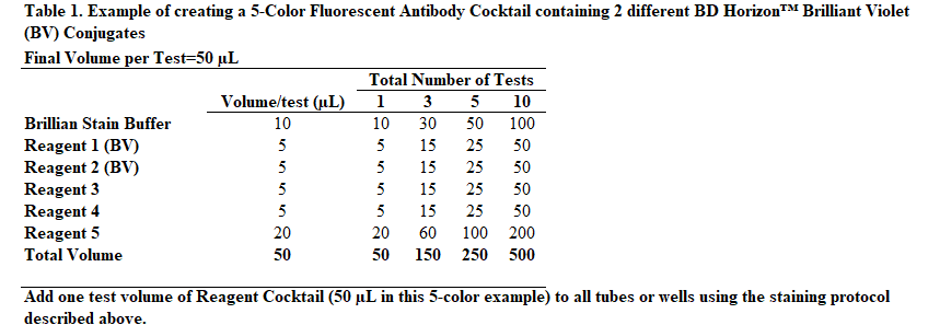
Instead of adding staining reagents individually to each tube or well of a 96-well plate, it may be desirable to add cocktailed staining reagents, ie, mixtures of two or more fluorescent staining reagents. The following protocol provides an example of how to prepare a "per test" 5-Color Fluorescent Antibody Cocktail that already contains BD Horizon Brilliant Stain Buffer Plus.
Human Samples: Pre-mixed Fluorescent Reagent Cocktails
For each multicolor test of cocktailed fluorescent reagents:
i) Add 10 μL of BD Horizon Brilliant Stain Buffer Plus per test
ii) Add each fluorescent reagent at the recommended volume per test (5 μL or 20 μL)
iii) Add BD Pharmingen Stain Buffer (FBS) (Cat. No. 554656) to bring the volume of each test to a minimum of 50 μL
iv) Mix reagents (especially after adding BD Horizon Brilliant reagents)
v) Store cocktail at 4°C protected from light if it is to be used later
Note: Protected from light, fluorescent reagent cocktails containing more than one Brilliant Violet and/or Brilliant Blue reagent are best used within 24 hours after preparation when stored at 4ºC or within 4 hours when stored at room temperature. However, when more than one Brilliant Ultraviolet (BUV) reagent is in the cocktail, it is best used within 2 hours after preparation irrespective of storage temperature.
For additional guidance, see Table 1 "Example of creating a 5-Color Fluorescent Antibody Cocktail containing 2 different BD Horizon™ Brilliant Violet (BV) Conjugates" at the end of this field.
Compensation and Setup
BD Horizon™ Brilliant Stain Buffer Plus can be used in single color compensation controls using cells. The buffer is compatible with BD® Compbeads, however, it has not been tested with compensation beads from other vendors. For the most accurate compensation, compensation controls created with either cells or beads should be exposed to BD Horizon™ Brilliant Stain Buffer for the same length of time as the corresponding multicolor panel. Additionally, the most accurate compensation will be created when BD Horizon™ Brilliant Stain Buffer is used in all compensation controls, including Brilliant polymer and non-polymer dyes.