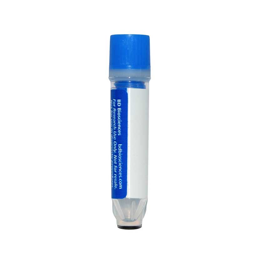
Preparation And Storage
Recommended Assay Procedures
Put all BD® AbSeq Reagents to be pooled into a Latch Rack for 500 µL Tubes (Thermo Fisher Scientific Cat. No. 4900). Arrange the tubes so that they can be easily uncapped and re-capped with an 8-Channel Screw Cap Tube Capper (Thermo Fisher Scientific Cat. No. 4105MAT) and the reagents aliquoted with a multi-channel pipette.
BD® AbSeq tubes should be centrifuged for ≥ 30 seconds at 400 × g to ensure removal of any content in the cap/tube threads prior to the first opening.
Product Notices
- This reagent has been pre-diluted for use at the recommended volume per test. Typical use is 2 µl for 1 × 10^6 cells in a 200-µl staining reaction.
- The production process underwent stringent testing and validation to assure that it generates a high-quality conjugate with consistent performance and specific binding activity. However, verification testing has not been performed on all conjugate lots.
- Please refer to bd.com/genomics-resources for technical protocols.
- Caution: Sodium azide yields highly toxic hydrazoic acid under acidic conditions. Dilute azide compounds in running water before discarding to avoid accumulation of potentially explosive deposits in plumbing.
- Source of all serum proteins is from USDA inspected abattoirs located in the United States.
- Illumina is a trademark of Illumina, Inc.
- Please refer to http://regdocs.bd.com to access safety data sheets (SDS).
- For U.S. patents that may apply, see bd.com/patents.
Companion Products


The NOK-1 monoclonal antibody specifically recognizes CD178. Fas (CD95; APO-1) is a 45 kDa cell surface protein that mediates apoptosis when cross-linked with agonistic anti-Fas antibodies or by Fas ligand (FasL; CD178). Fas belongs to the TNF (Tumor Necrosis Factor)/NGF (Nerve Growth Factor) receptor family, and is expressed in various tissues and cells including the thymus, liver, ovary and lung. CD178 (FasL), a member of the TNF cytokine family, induces apoptosis by binding to Fas, its cell-surface receptor. FasL may exist as either membrane bound or soluble forms and is expressed by activated T and NK cells. FasL may also be constitutively expressed in some immunologically privileged sites, e.g., eye and testis. Fas and FasL play an important role in the induction of apoptosis, and thus regulate a variety of immunological responses.
The NOK-1 antibody clone has been reported to recognize human FasL, recognizing both the membrane bound (FasL) and soluble (sFasL) forms. It is reported that the epitope for NOK-1 has been mapped to the COOH-terminus of FasL, at the region implicated in Fas binding. FasL and sFasL have been reported to migrate at reduced molecular weights of 40 and 26 kDa, respectively. However, the molecular weights observed in a particular sample may vary according to FasL and sFasL glycosylation and breakdown patterns as described in the literature. The NOK-1 antibody clone is not recommended for the Western blot application.
Development References (9)
-
Kayagaki N, Kawasaki A, Ebata T, et al. Metalloproteinase-mediated release of human Fas ligand. J Exp Med. 1995; 182(6):1777-1783. (Immunogen: Flow cytometry, Neutralization). View Reference
-
Orlinick JR, Elkon KB, Chao MV. Separate domains of the human fas ligand dictate self-association and receptor binding. J Biol Chem. 1997; 272(51):32221-32229. (Clone-specific: Flow cytometry). View Reference
-
Oyaizu N, Adachi Y, Hashimoto F, et al. Monocytes express Fas ligand upon CD4 cross-linking and induce CD4+ T cells apoptosis: a possible mechanism of bystander cell death in HIV infection. J Immunol. 1997; 158(5):2456-2463. (Clone-specific: Flow cytometry). View Reference
-
Sieg S, Smith D, Yildirim Z, Kaplan D. Fas ligand deficiency in HIV disease. Proc Natl Acad Sci U S A. 1997; 94(11):5860-5865. (Clone-specific: Flow cytometry). View Reference
-
Takahashi T, Tanaka M, Brannan CI, et al. Generalized lymphoproliferative disease in mice, caused by a point mutation in the Fas ligand. Cell. 1994; 76(6):969-976. (Biology). View Reference
-
Tanaka M, Suda T, Takahashi T, Nagata S. Expression of the functional soluble form of human Fas ligand in activated lymphocytes. EMBO J. 1995; 14(6):1129-1135. (Biology). View Reference
-
Villunger A, Egle A, Marschitz I, et al. Constitutive expression of Fas (Apo-1/CD95) ligand on multiple myeloma cells: a potential mechanism of tumor-induced suppression of immune surveillance. Blood. 1997; 90(1):12-20. (Clone-specific: Flow cytometry, Neutralization). View Reference
-
Walker PR, Saas P, Dietrich PY. Role of Fas ligand (CD95L) in immune escape: the tumor cell strikes back. J Immunol. 1997; 158(10):4521-4524. (Clone-specific: Neutralization). View Reference
-
Zavazava N, Kronke M. Soluble HLA class I molecules induce apoptosis in alloreactive cytotoxic T lymphocytes. Nat Med. 1996; 2(9):1005-1010. (Clone-specific: Flow cytometry, Neutralization). View Reference
Please refer to Support Documents for Quality Certificates
Global - Refer to manufacturer's instructions for use and related User Manuals and Technical data sheets before using this products as described
Comparisons, where applicable, are made against older BD Technology, manual methods or are general performance claims. Comparisons are not made against non-BD technologies, unless otherwise noted.
For Research Use Only. Not for use in diagnostic or therapeutic procedures.