Skip to main content Skip to navigation
Mouse T Lymphocyte Enrichment Set-DM

BD IMag™ Mouse T Lymphocyte Enrichment Set-DM

(RUO)
Mouse T Lymphocyte Enrichment Set-DM
Enrichment of T lymphocytes from mouse spleen. BALB/c splenocytes were labeled with the BD IMag™ Mouse T Lymphocyte Enrichment Set - DM (Cat. No. 557793) and separated on the BD IMag™ Cell Separation Magnet (Cat. No. 552311) according to the accompanying protocol. To demonstrate the efficiency of the enrichment, cells were stained with FITC Hamster Anti-Mouse CD3e (Cat. No. 553061/553062) to detect T lymphocytes and a mixture of PE Anti-Mouse CD11b (Cat. No. 557397/553311), Anti-Mouse CD45/B220 (Cat. No. 553089/553090), Anti-Mouse 49b (Cat. No. 558759), and Anti-Mouse Ter-119/Erythroid Cells (Cat. No. 553673) monoclonal antibodies to detect non-T leukocytes and erythrocytes. Dead cells were excluded by staining with Propidium Iodide Staining Solution (Cat. No. 556463). Flow cytometry was performed on a BD FACSCalibur™ flow cytometry system. Please refer to the Enrichment Flow Chart on the next page to identify the cell populations represented in this figure. The percentage of T cells is indicated in the lower-right corner of each panel. The far left panel shows unseparated splenocytes.  The middle left panel shows the combined enriched fraction after three 6-minute magnetic separations. The middle right panel shows the twice-enriched fraction after an additional 6-minute separation of the cells shown in the middle left panel. This additional incubation can result in up to 5% increased purity with up to a 5% decrease in recovery.  The far right panel shows the positive fraction.
Product Details
Down Arrow Up Arrow


BD IMag™
Cell separation (Routinely Tested)
RUO
AB_2869096


Description

The BD IMag™ Mouse T Lymphocyte Enrichment Set - DM is used for the negative selection of T lymphocytes from mouse spleen or lymph node. The Biotinylated Mouse T Lymphocyte Enrichment Cocktail contains monoclonal antibodies that recognize antigens expressed on peripheral erythrocytes and leukocytes that are not T lymphocytes. The BD IMag™ Streptavidin Particles Plus - DM are magnetic nanoparticles that have streptavidin covalently conjugated to their surfaces. With these two components, the BD IMag™ Mouse T Lymphocyte Enrichment Set -DM avoids the inadvertent activation of the enriched T lymphocytes by using reagents that do not directly bind to those T cells. This Set has been optimized for use with the BD IMag™ Cell Separation Magnet, and it contains sufficient reagents to label 10^9 leukocytes.  

The Biotinylated Mouse T Lymphocyte Enrichment Cocktail is comprised of the following biotin-conjugated monoclonal antibodies:

Anti-mouse CD11b, clone M1/70

Anti-mouse CD45R/B220, clone RA3-6B2

Anti-mouse CD49b, clone HMα2

Anti-mouse TER-119/Erythroid Cells, clone TER-119

Preparation And Storage

Store undiluted at 4°C. The monoclonal antibody was purified from tissue culture supernatant or ascites by affinity chromatography. The antibody was conjugated with biotin under optimum conditions, and unreacted biotin was removed. Antibody or streptavidin was conjugated to the magnetic particles under optimum conditions, and unconjugated antibody/streptavidin was removed.

Recommended Assay Procedures

The detailed Magnetic Labeling and Enrichment Protocol follows. In summary, the Biotinylated Mouse T Lymphocyte Enrichment Cocktail simultaneously stains erythrocytes and most leukocytes except the T lymphocytes. After washing away excess antibody, BD IMag™ Streptavidin Particles Plus - DM are added to the cell suspension and bind the cells bearing the biotinylated antibodies. The tube containing this labeled cell suspension is then placed within the magnetic field of the BD IMag™ Cell Separation Magnet (Cat. No. 552311). Negative selection is then performed to enrich for the unlabeled T cells. Labeled cells migrate toward the magnet (positive fraction), leaving the unlabeled cells in suspension so they can be drawn off and retained (enriched fraction). The negative selection is repeated twice to increase the yield of the enriched fraction. If greater purity is required, negative selection may be performed on the enriched fraction. The magnetic separation steps are diagrammed in the Enrichment Flow Chart. The positive and enriched fractions can be evaluated in downstream applications such as flow cytometry and tissue culture. The antibodies in the Biotinylated Mouse T Lymphocyte Enrichment Cocktail have been optimized and pre-diluted to maximize efficiency of T lymphocyte enrichment from peripheral lymphoid organs.

MAGNETIC LABELING AND ENRICHMENT PROTOCOL

1.   All labeling and enrichment steps may be performed in either tissue culture medium* or sterile 1X BD IMag™ buffer.

- For 1X BD IMag™ buffer: Dilute BD IMag™ Buffer (10X) (Cat. No. 552362) 1:10 with sterile distilled water or prepare Phosphate Buffered Saline supplemented with 0.5% BSA, 2 mM EDTA, and 0.1% sodium azide.

2.   Aseptically prepare a single-cell suspension from the peripheral lymphoid tissue of interest. Remove clumps of cells and/or debris by passing the suspended cells through a 70-µm nylon cell strainer. Cell suspensions can be prepared in tissue culture medium* or 1X BD IMag™ buffer.

3.   Count the cells. If the concentration is between 10x10^6 and 20x10^6 cells/ml, proceed to Step 4. If cells are more dilute, spin down the cells and resuspend them in tissue culture medium* or 1X BD IMag™ buffer at a concentration of 20 x 10^6 cells/ml.

4.   Add the Bioinylated Mouse T Lymphocyte Enrichment Cocktail at 5 µl per 1 x 10^6 cells, and incubate on ice for 15 minutes.†

5.   Wash the labeled cells with a 10X excess volume of tissue culture medium* or 1X BD IMag™ buffer, centrifuge at 300 x g for 7 minutes, and carefully aspirate ALL the supernatant.

6.   Vortex the BD IMag™ Streptavidin Particles Plus - DM thoroughly, and add 5 µl of particles for every 1 x 10^6 total cells.

7.   MIX THOROUGHLY. Refrigerate for 30 minutes at 6°C - 12°C.†

8.   Bring the labeling concentration up to 20-80 x 10^6 cells/ml with tissue culture medium* or 1X BD IMag™ buffer.

9.   Transfer the labeled cells to a 12 x 75 mm round-bottom test tube, maximum volume added not to exceed 1.0 ml. Place this positive-fraction tube on the Cell Separation Magnet (horizontal position) for 6 to 8 minutes.†

- For greater volume, transfer the cells to a 17 x 100 mm round-bottom test tube, maximum volume added not to exceed 3.0 ml. Place this positive-fraction tube on the Cell Separation Magnet (vertical position) for 8 minutes.†

10.  With the tube on the Cell Separation Magnet and using a sterile glass Pasteur pipette, carefully aspirate the supernatant (enriched fraction) and place in a new sterile tube.

11.  Remove the positive-fraction tube from the Cell Separation Magnet, and add tissue culture medium* or 1X BD IMag™ buffer to the same volume as in Step 8. Resuspend the positive fraction well by pipetting up and down 10 to 15 times, and place the tube back on the Cell Separation Magnet for 6 to 8 minutes.†

- For 17 x 100 mm tube: Place on the Cell Separation Magnet for 8 minutes.†

12.  Using a new sterile Pasteur pipette, carefully aspirate the supernatant and combine with the enriched fraction from Step 10 above.

13.  Repeat Steps 11 and 12. The combined enriched fraction contains T lymphocytes with no bound antibodies or magnetic particles. These cells are ready for downstream applications, or they can be further enriched by proceeding to Step 15.

14.  The positive-fraction cells remaining in the original tube can be resuspended in an appropriate buffer or culture medium for downstream applications, including flow cytometry, if desired.

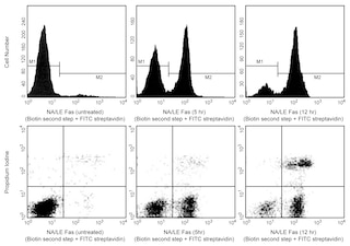
15.  To increase the purity of the combined enriched fraction by another 3% to 5% (compare the middle left and middle right panels in the figure), place the tube containing the combined enriched fraction on the Cell Separation Magnet for another 6 to 8 minutes.†

- For 17 x 100 mm tube: Place on the Cell Separation Magnet for 8 minutes.†

16.  Carefully aspirate the supernatant and place in a new sterile tube.  This is the twice-enriched fraction. The cells are ready to be processed for downstream applications.

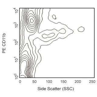
17.  Samples of the total cell suspension and the positive and enriched fractions should be analyzed by flow cytometry to evaluate the efficiency of the cell-separation procedure.

NOTES: * Some tissue culture media contain biotin, which may interfere with the binding of the Streptavidin Particles. We recommend Dulbecco's Minimum Essential Medium (DMEM).

† Avoid nonspecific labeling by working quickly and adhering to recommended incubation times.

Product Notices

  1. BD IMag™ particles are prepared from carboxy-functionalized magnetic particles which are manufactured by Skold Technology and are licensed under US patent number 7,169,618.
  2. Caution: Sodium azide yields highly toxic hydrazoic acid under acidic conditions. Dilute azide compounds in running water before discarding to avoid accumulation of potentially explosive deposits in plumbing.
  3. Please refer to www.bdbiosciences.com/us/s/resources for technical protocols.
  4. Since applications vary, each investigator should titrate the reagent to obtain optimal results.
  5. Source of all serum proteins is from USDA inspected abattoirs located in the United States.
557793 Rev. 2
Components
Down Arrow Up Arrow
Description EntrezGene ID
Biotinylated Mouse T Lymphocyte Enrichment Cocktail N/A
Streptavidin Particles Plus - DM N/A
557793 Rev. 2

Please refer to Support Documents for Quality Certificates

Global - Refer to manufacturer's instructions for use and related User Manuals and Technical data sheets before using this products as described

Comparisons, where applicable, are made against older BD Technology, manual methods or are general performance claims.  Comparisons are not made against non-BD technologies, unless otherwise noted.

For Research Use Only. Not for use in diagnostic or therapeutic procedures.