Immune cell functions in homeostasis and disease
Interview with Florian Mair, Research Associate, Fred Hutchinson Cancer Research Center, USA
October 28, 2021

In our continuing coverage of scientists working at the frontiers of immunology, we are pleased to present an interview with Florian Mair, a Research Associate at the Prlic Lab, Fred Hutchinson Cancer Research Center, USA. Florian has been working on distinguishing human tumor-unique alterations from non-malignant tissue inflammation. In this interview, he talked to us about his work, the role of novel technologies and his vision for the future of immunology.
1. Tell us something about yourself and your scientific interests.
My main interests are how immune cell function is regulated during immune homeostasis and disease in human tissues. Within the many different immune cell populations that exist, I am mostly interested in the interplay between dendritic cells (DCs) and effector and regulatory T cells. Also, I enjoy leveraging novel technologies and computational data analysis approaches to explore the amazing complexity of these cells in more detail.
2. Can you give us a bit of history as to how the immune processes in inflamed tissues were found to be relevant for tumors? What can we learn about the tumor microenvironment from inflammation?
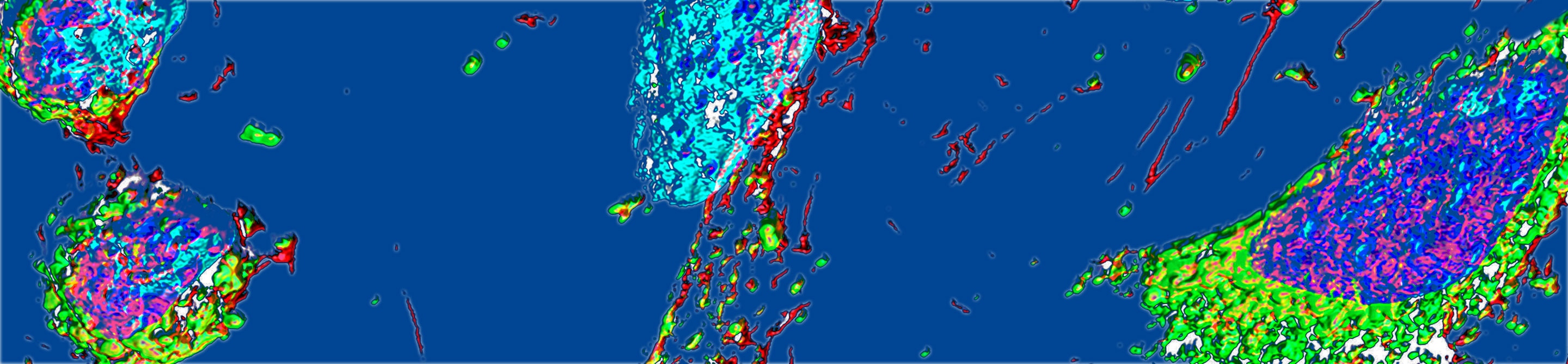
Over the past years, it has become evident that immune cells found in the blood are not a good representation of what is going on in solid tissues, which is where the actual effector immune response is happening. Our lab has been interested in inflammatory processes in human tissues for a while, and over time we discovered that the immune signatures in these samples were surprisingly similar to what has been reported for human tumors. That’s when we started wondering whether we could leverage this comparison to identify immune cell subsets that are truly unique to the tumor microenvironment and not just a result of general inflammatory processes.
3. Can you tell us something about the insights that can be obtained by combining flow cytometry and other techniques like multi-omics in single-cell biology?
When working with human tissue samples, these are almost always very limited in size. Thus, we must utilise techniques where we can gain as much insight as possible from a limited number of cells. Combining high-dimensional flow cytometry with multi-omic single-cell RNA sequencing (scRNA-seq) is currently one of the best approaches to achieve this. For example, one advantage of multi-omic scRNA-seq is that one can perform “unbiased” clustering based on transcript expression, and then map the resulting cellular clusters back to a particular surface protein phenotype, which can be used to design downstream sorting experiments.
4. In your recent webinar, you presented the RNA sequence analysis as the final step. How can you follow up on those findings?
As mentioned above, I think an exploratory analysis of transcriptomes is only one step in the process. We as a scientific community need to do follow-up studies, where newly identified cellular phenotypes are probed ex vivo in additional experiments, for example, stimulation with cytokines, TCR signals, etc.
5. What is your vision for the future of immunology? Can you tell us something about the direction immunology research will take – the hot topics, trends, etc?
I think one hot topic is advancing all these high-dimensional techniques into a spatial dimension, i.e. be able to directly look at the transcriptome and surface protein phenotype of cells inside tissues without the need for isolation. Also, I think as the cost for high-dimensional single-cell analysis continues to fall, there are potentially huge opportunities in the context of personalized medicine and immunomonitoring. For all of this, we need advanced (yet easy-to-use) computational analysis approaches that help us make sense of these complex data.
The views expressed by the interviewee are his personal opinions.