Old Browser
This page has been recently translated and is available in French now.
Looks like you're visiting us from {countryName}.
Would you like to stay on the current country site or be switched to your country?


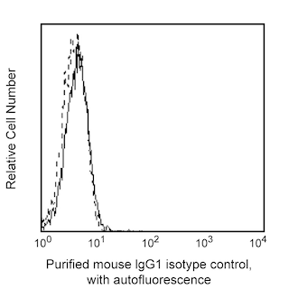
Flow cytometric analysis of CD133 expression on human retinoblastoma cell line. Cells from the WERI-Rb-1 (Retinoblastoma, ATCC HTB-169) cell line were stained with either Purified Mouse IgG1, κ Isotype Control (Cat. No. 556648; dotted line histogram) or Purified Mouse Anti-Human CD133 antibody (Cat. No. 567210; solid line histogram) at 0.125 μg/test. The cells were washed and stained with PE Goat Anti-Mouse Ig (Cat. No. 550589). The fluorescence histogram showing CD133 expression (or Ig Isotype control staining) was derived from gated events with the forward and side light-scatter characteristics of viable cells. Flow cytometry and data analysis were performed using a BD LSRFortessa™ Cell Analyzer System and FlowJo™ software. Data shown on this Technical Data Sheet are not lot specific.

Immunohistochemical analysis of human CD133 expression by cells comprising human placenta tissue. Cold acetone-fixed frozen sections of human placenta were stained with either Purified Mouse IgG1, κ Isotype Control (Cat. No. 550878; Left Panel) or Purified Mouse Anti-Human CD133 antibody (Cat. No. 567210; Right Panel) at 5 µg/ml for one hour at room temperature. A three-step staining procedure that employs a Biotin Goat Anti-Mouse Ig (Multiple Adsorption) (Cat. No. 550337), Streptavidin-Horseradish Peroxidase (Sav-HRP) (Cat. No. 550946), and the DAB Substrate Kit (Cat. No. 550880) was used to develop the primary staining reagents. The tissues were counterstained with hematoxylin. CD133 positive staining on syncytiotrophoblasts of chorionic villi (Right Panel) is indicated by the intense brown labeling. Original magnification: 20X. Data shown on this Technical Data Sheet are not lot specific.


BD Pharmingen™ Purified Mouse Anti-Human CD133

BD Pharmingen™ Purified Mouse Anti-Human CD133

Regulatory Status Legend
Any use of products other than the permitted use without the express written authorization of Becton, Dickinson and Company is strictly prohibited.
Preparation And Storage
Product Notices
- Since applications vary, each investigator should titrate the reagent to obtain optimal results.
- An isotype control should be used at the same concentration as the antibody of interest.
- Caution: Sodium azide yields highly toxic hydrazoic acid under acidic conditions. Dilute azide compounds in running water before discarding to avoid accumulation of potentially explosive deposits in plumbing.
- Sodium azide is a reversible inhibitor of oxidative metabolism; therefore, antibody preparations containing this preservative agent must not be used in cell cultures nor injected into animals. Sodium azide may be removed by washing stained cells or plate-bound antibody or dialyzing soluble antibody in sodium azide-free buffer. Since endotoxin may also affect the results of functional studies, we recommend the NA/LE (No Azide/Low Endotoxin) antibody format, if available, for in vitro and in vivo use.
- Please refer to http://regdocs.bd.com to access safety data sheets (SDS).
- Please refer to www.bdbiosciences.com/us/s/resources for technical protocols.
Companion Products


.png?imwidth=320)
The W6B3C1 monoclonal antibody specifically recognizes CD133 which is also known as Prominin-like protein 1 (PROML1), Prominin-1 (PROM1), hProminin, Hematopoietic stem cell antigen, Macular dystrophy retinal 2 (MCDR2), Stargardt disease 4 autosomal dominant (STGD4), or AC133 antigen. CD133 is an ~120 kDa five-transmembrane, glycoprotein that is encoded by PROM1 (Prominin 1) which belongs to the Prominin gene family. This single-chain, pentaspan transmembrane glycoprotein is comprised of an extracellular N-terminus with two short intracellular sequences and two long extracellular loops followed by an intracellular C-terminus. CD133 is expressed on some cells found in a variety of tissues including the bone marrow, cord and peripheral blood, placenta, liver, pancreas, kidney, lung, retina, brain and heart. It is expressed on various cell types including hematopoietic stem cells and progenitor cells, neural stem cells, developing epithelial cells, precursor endothelial cells, and retinal cells. CD133 is expressed on some cancer cells found in leukemias, melanoma and retinoblastoma. It may serve as a cancer stem cell marker in a number of brain tumors, melanoma, colon cancer, hepatocellular carcinoma, pancreatic adenocarcinoma, and prostate cancer. A mutation in PROM1 is reportedly associated with a form of human retinal degeneration. The W6B3C1 antibody recognizes a different epitope than the human CD133-specific 293C3 antibody.
Development References (7)
-
Breznik B, Limbaeck Stokin C, Kos J, et al. Cysteine cathepsins B, X and K expression in peri-arteriolar glioblastoma stem cell niches.. J Mol Histol. 2018; 49(5):481-497. (Clone-specific: Immunohistochemistry). View Reference
-
Bühring HK, Marzer A, Lammers R, Wissinger B. CD133 cluster report. In: Mason D. David Mason .. et al., ed. Leucocyte typing VII : white cell differentiation antigens : proceedings of the Seventh International Workshop and Conference held in Harrogate, United Kingdom. Oxford: Oxford University Press; 2002:622-623.
-
Koerner SP, André MC, Leibold JS, et al. An Fc-optimized CD133 antibody for induction of NK cell reactivity against myeloid leukemia.. Leukemia. 2017; 31(2):459-469. (Clone-specific: Blocking, Flow cytometry). View Reference
-
Kong DS, Kim MH, Park WY, et al. The progression of gliomas is associated with cancer stem cell phenotype.. Oncol Rep. 2008; 19(3):639-43. (Clone-specific: Immunohistochemistry). View Reference
-
Lammers R, Giesert C, Grünebach F, Marxer A, Vogel W, Bühring HJ. Monoclonal antibody 9C4 recognizes epithelial cellular adhesion molecule, a cell surface antigen expressed in early steps of erythropoiesis.. Exp Hematol. 2002; 30(6):537-45. (Immunogen: Flow cytometry). View Reference
-
Maw MA, Corbeil D, Koch J, et al. A frameshift mutation in prominin (mouse)-like 1 causes human retinal degeneration.. Hum Mol Genet. 2000; 9(1):27-34. (Biology). View Reference
-
Miraglia SJ, Buck D. CD133 (AC133). In: Mason D. David Mason .. et al., ed. Leucocyte typing VII : white cell differentiation antigens : proceedings of the Seventh International Workshop and Conference held in Harrogate, United Kingdom. Oxford: Oxford University Press; 2002:870-872.
Please refer to Support Documents for Quality Certificates
Global - Refer to manufacturer's instructions for use and related User Manuals and Technical data sheets before using this products as described
Comparisons, where applicable, are made against older BD Technology, manual methods or are general performance claims. Comparisons are not made against non-BD technologies, unless otherwise noted.
For Research Use Only. Not for use in diagnostic or therapeutic procedures.