





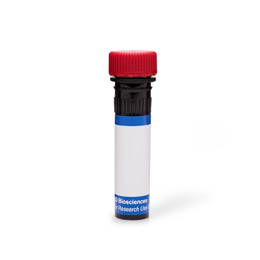
BD Pharmingen™ PE Rat Anti-Mouse CD131
Clone JORO50 (also known as JORO 50) (RUO)


Flow cytometric analysis of βIL-3 and CD131 expression on Mac-1+ bone marrow cells. BALB/c bone marrow cells (BMC) were stained (20 minutes at 4°C) with PE Rat Anti-Mouse CD131 antibody (1 µg mAb/10^6 cells; Cat. No. 559920) and FITC Rat Anti-Mouse CD11b (Cat. No. 553310). The two-color fluorescence dot plot shows the correlated expression patterns of CD11b versus βIL-3 and CD131 with the forward and side light-scatter characteristics of viable bone marrow cells.
Flow cytometric analysis of βIL-3 and CD131 expression on Mac-1+ bone marrow cells. BALB/c bone marrow cells (BMC) were stained (20 minutes at 4°C) with PE Rat Anti-Mouse CD131 antibody (1 µg mAb/10^6 cells; Cat. No. 559920) and FITC Rat Anti-Mouse CD11b (Cat. No. 553310). The two-color fluorescence dot plot shows the correlated expression patterns of CD11b versus βIL-3 and CD131 with the forward and side light-scatter characteristics of viable bone marrow cells.


Preparation And Storage


Product Notices
- Since applications vary, each investigator should titrate the reagent to obtain optimal results.
- An isotype control should be used at the same concentration as the antibody of interest.
- Caution: Sodium azide yields highly toxic hydrazoic acid under acidic conditions. Dilute azide compounds in running water before discarding to avoid accumulation of potentially explosive deposits in plumbing.
- For fluorochrome spectra and suitable instrument settings, please refer to our Multicolor Flow Cytometry web page at www.bdbiosciences.com/colors.
- Please refer to www.bdbiosciences.com/us/s/resources for technical protocols.
Companion Products


The JORO50 monoclonal antibody specifically recognizes the extracellular regions of the mouse CD131/βc (Bc, AIC2B, Il3rb1, Csf2rb1) and βIL3 (BIL3, AIC2A, Il3rb2, Csf2rb2) cytokine receptor subunits. A variety of mouse cell types, including multipotential hematopoietic stem cells, mast cells, megakaryocytes, eosinophils, erythroblasts, pre-B cells, and osteoclasts, express βIL-3 and βc subunits. Either βIL-3 or βc can combine with the IL-3Rα chain to form two distinct, functional (i.e., signaling), high affinity receptors for mouse IL-3. The βIL-3 subunit by itself can bind mouse IL-3 with low affinity whereas the βc subunit can not. βc (but not βIL-3) can combine with the IL-5Rα (CD125) and GM-CSFRα (CD116) subunits to form high affinity, signaling receptors for mouse IL-5 or GM-CSF, respectively. The immunogen used to generate the JORO50 hybridoma was the bone marrow-derived, C4-77 pro-T lymphocyte clone.


No Citations Are Available for this Product
Please refer to Support Documents for Quality Certificates
Global - Refer to manufacturer's instructions for use and related User Manuals and Technical data sheets before using this products as described
Comparisons, where applicable, are made against older BD Technology, manual methods or are general performance claims. Comparisons are not made against non-BD technologies, unless otherwise noted.
For Research Use Only. Not for use in diagnostic or therapeutic procedures.