-
Reagents
- Flow Cytometry Reagents
-
Western Blotting and Molecular Reagents
- Immunoassay Reagents
-
Single-Cell Multiomics Reagents
- BD® OMICS-Guard Sample Preservation Buffer
- BD® AbSeq Assay
- BD® Single-Cell Multiplexing Kit
- BD Rhapsody™ ATAC-Seq Assays
- BD Rhapsody™ Whole Transcriptome Analysis (WTA) Amplification Kit
- BD Rhapsody™ TCR/BCR Next Multiomic Assays
- BD Rhapsody™ Targeted mRNA Kits
- BD Rhapsody™ Accessory Kits
- BD® OMICS-One Protein Panels
- BD OMICS-One™ WTA Next Assay
-
Functional Assays
-
Microscopy and Imaging Reagents
-
Cell Preparation and Separation Reagents
Old Browser
This page has been recently translated and is available in French now.
Looks like you're visiting us from {countryName}.
Would you like to stay on the current location site or be switched to your location?
BD Pharmingen™ Alexa Fluor® 488 Mouse anti-Human TRA-1-81 Antigen
Clone TRA-1-81 (RUO)

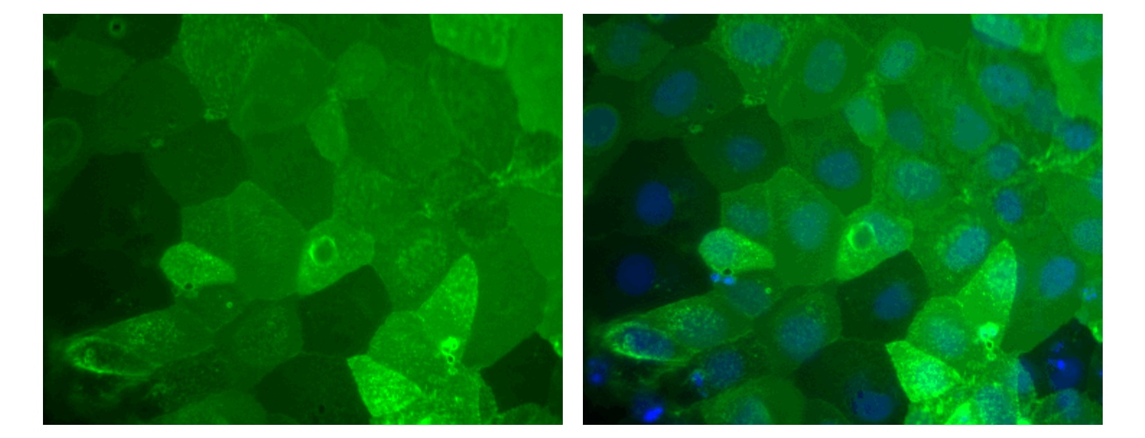
Immunofluorescent staining of human ES cell line. The H9 cell line (WiCell, Madison, WI) was cultured, fixed, and stained with Alexa Fluor® 488 Mouse anti-Human TRA-1-81 Antigen monoclonal antibody (pseudo-colored green) according to the Recommended Assay Procedure. The left image shows the plasma membrane staining by the TRA-1-81 mAb, and the right image shows TRA-1-81 with counter-staining of the nuclei by Hoechst 33342 (pseudo-colored blue). The images were captured on a BD Pathway™ 435 Cell Analyzer using a 10X objective and merged using BD Attovision™ software.


Immunofluorescent staining of human ES cell line. The H9 cell line (WiCell, Madison, WI) was cultured, fixed, and stained with Alexa Fluor® 488 Mouse anti-Human TRA-1-81 Antigen monoclonal antibody (pseudo-colored green) according to the Recommended Assay Procedure. The left image shows the plasma membrane staining by the TRA-1-81 mAb, and the right image shows TRA-1-81 with counter-staining of the nuclei by Hoechst 33342 (pseudo-colored blue). The images were captured on a BD Pathway™ 435 Cell Analyzer using a 10X objective and merged using BD Attovision™ software.

Immunofluorescent staining of human ES cell line. The H9 cell line (WiCell, Madison, WI) was cultured, fixed, and stained with Alexa Fluor® 488 Mouse anti-Human TRA-1-81 Antigen monoclonal antibody (pseudo-colored green) according to the Recommended Assay Procedure. The left image shows the plasma membrane staining by the TRA-1-81 mAb, and the right image shows TRA-1-81 with counter-staining of the nuclei by Hoechst 33342 (pseudo-colored blue). The images were captured on a BD Pathway™ 435 Cell Analyzer using a 10X objective and merged using BD Attovision™ software.


Regulatory Status Legend
Any use of products other than the permitted use without the express written authorization of Becton, Dickinson and Company is strictly prohibited.
Preparation And Storage
Recommended Assay Procedures
1. Seed the cells in appropriate culture medium at an appropriate cell density in a BD Falcon™ 96-well Imaging Plate (Cat. No. 353219), and
culture overnight to 48 hours.
2. Remove the culture medium from the wells, wash the wells twice with 100 μl of 1× PBS, and fix the cells by adding 100 µl of fresh 3.7% Formaldehyde in PBS or BD Cytofix™ fixation buffer (Cat. No. 554655) to each well and incubating for 10 minutes at room temperature (RT).
3. Remove the fixative from the wells, and wash the wells twice with 100 μl of 1× PBS.
4. Dilute the antibody 1:10 in 1× PBS, and stain the cells by adding 50 µl of the diluted antibody conjugate to each well and incubating for 1 hour at RT.
5. Remove the diluted antibody, and wash the wells twice with 100 μl of 1× PBS.
6. Remove the PBS, and counter-stain the nuclei by adding 100 μl of a 2 μg/ml solution of Hoechst 33342 (eg, Sigma-Aldrich Cat. No. B2261) in 1× PBS to each well at least 15 minutes before imaging.
7. View and analyze the cells on an appropriate imaging instrument. Recommended filters for the BD Pathway™ cell analyzers are:
Instrument Excitation Emission Dichroic
BD Pathway 855 488/10 515 LP Fura/FITC
BD Pathway 435 482/35 536/40 FF506
Product Notices
- Please refer to www.bdbiosciences.com/us/s/resources for technical protocols.
- This reagent has been pre-diluted for use at the recommended Volume per Test when following the Recommended Assay Procedure. A Test is typically ~10,000 cells cultured in a well of a 96-well imaging plate.
- The Alexa Fluor®, Pacific Blue™, and Cascade Blue® dye antibody conjugates in this product are sold under license from Molecular Probes, Inc. for research use only, excluding use in combination with microarrays, or as analyte specific reagents. The Alexa Fluor® dyes (except for Alexa Fluor® 430), Pacific Blue™ dye, and Cascade Blue® dye are covered by pending and issued patents.
- Source of all serum proteins is from USDA inspected abattoirs located in the United States.
- Caution: Sodium azide yields highly toxic hydrazoic acid under acidic conditions. Dilute azide compounds in running water before discarding to avoid accumulation of potentially explosive deposits in plumbing.
- Alexa Fluor® is a registered trademark of Molecular Probes, Inc., Eugene, OR.
The TRA-1-81 monoclonal antibody reacts with a pluripotent stem cell-specific epitope on a high molecular weight transmembrane glycoprotein. The TRA-1-81 antigen is an epitope on the same keratan sulfate core molecule, podocalyxin, as 4 other distinct antigens on tumor-derived cell lines, TRA-1-60, GCTM2, K4, and K21. The expression of TRA-1-81 antigen is stage-specific and can be used to characterize embryonic cells and monitor their differentiation. The antigen is found on teratocarcinoma (embryonal carcinoma or EC), embryonic inner cell mass (but not morula or trophoblast), and embryonic stem (ES) cells. As human EC and ES cells undergo differentiation, expression of TRA-1-81 antigen is lost.

Development References (7)
-
Andrews PW, Banting G, Damanov I, Arnaud D, Avner P. Three monoclonal antibodies defining distinct differentiation antigens associated with different high molecular weight polypeptides on the surface of human embryonal carcinoma cells. Hybridoma. 1984; 3(4):347-361. (Immunogen: Immunofluorescence, Immunoprecipitation, Radioimmunoassay). View Reference
-
Badcock G, Pigott C, Goepel J, Andrews PW. The human embryonal carcinoma marker antigen TRA-1-60 is a sialylated keratan sulfate proteoglycan. Cancer Res. 1999; 59:4715-4719. (Clone-specific: Immunoprecipitation, Western blot). View Reference
-
Draper JS, Pigott C, Thomson JA, Andrews PW. Surface antigens of human embryonic stem cells: changes upon differentiation in culture. J Anat. 2002; 200:249-258. (Clone-specific: Flow cytometry). View Reference
-
Henderson JK, Draper JS, Baillie HS, et al. Preimplantation human embryos and embryonic stem cells show comparable expression of stage-specific embryonic antigens. Stem Cells. 2002; 20:329-337. (Clone-specific: Flow cytometry, Immunofluorescence). View Reference
-
Schopperle WM, DeWolf WC. The TRA-1-60 and TRA-1-81 human pluripotent stem cell markers are expressed on podocalyxin in embryonal carcinoma. Stem Cells. 2007; 25:723-730. (Clone-specific: Immunofluorescence, Western blot). View Reference
-
Thomson JA, Itskovitz-Eldor J, Shapiro SS, et al. Embryonic stem cell lines derived from human blastocysts. Science. 1998; 282:1145-1147. (Clone-specific: Immunocytochemistry (cytospins)). View Reference
-
Thomson JA, Kalishman J, Golos TG, et al. Isolation of a primate embryonic stem cell line. Proc Natl Acad Sci U S A. 1995; 92:7844-7848. (Clone-specific: Immunocytochemistry (cytospins)). View Reference
Please refer to Support Documents for Quality Certificates
Global - Refer to manufacturer's instructions for use and related User Manuals and Technical data sheets before using this products as described
Comparisons, where applicable, are made against older BD Technology, manual methods or are general performance claims. Comparisons are not made against non-BD technologies, unless otherwise noted.
For Research Use Only. Not for use in diagnostic or therapeutic procedures.

