Fluorochrome Faceoff Series: Performance Data Determine the Best Fluorochromes for Flow Cytometry Panel Design
June 30, 2025
This past fall, scientists had the opportunity to root for their favorite fluorochrome in the Fluorochrome Faceoff Series—a playful yet fierce competition that pitted flow cytometry fluorochromes against each other for medals and bragging rights across six different categories. Scientists voted for their favorite dyes, but the winning dyes were awarded based on performance.
The Fluorochrome Faceoff games were designed around the expected performance of high-quality dyes. For example, one game focused on which fluorochrome provided the highest resolution, while another addressed cross-laser excitation (spillover). Fluorochrome photostability and intracellular performance were also part of the fun. The contenders in this competition were an assortment of fluorochromes excited primarily by blue and yellow-green lasers.
Scientists from all over the world submitted their votes for each category. The performance of these blue and yellow-green dyes was evaluated using flow cytometry data, and the winners were announced on a bi-weekly basis. The scientists' votes were well aligned with the actual results of the competition.
If you missed the excitement, here's a summary of the competition and its results:
Fluorochrome Evaluation Categories
Several characteristics were evaluated and the fluorochromes were ranked based on how they performed. Specifically, the categories in which the dyes were evaluated were:
Competitors: Fluorochromes that were entered into this contest ranged from veterans such as PE and PerCP tandem dyes like PerCP-Cy5.5 and PE-Cy7 to newly available fluorochromes such as the BD Horizon RealYellow™ and RealBlue™ Dyes, NovaFluor™ Dyes and StarBright™ Dyes.
Competition: Category descriptions were presented along with experimental analysis to support the results. Winning medals were assigned to the best performing dyes in each category and runner-up dyes were also acknowledged for their performance.
Results
Overall Summary: The number of winners varied across the categories. In some games, like Resolution, almost all participants were winners, with three out of four fluorochromes selected as winners and the fourth as a runner-up. However, in the Spillover game, only two of six fluorochromes were winners, with one runner-up. For the Intracellular game, all tested dyes were winners, but many dyes were not available for intracellular markers and thus did not participate.
Key takeaway: BD Horizon RealYellow™ and RealBlue™ Dyes achieved a winning ranking in every faceoff, whereas other fluorochrome options had sporadic successes, winning in one or two instances but not even placing in others.
| Dye Family | Spillover | Resolution | Buffer Compatibility | Intracellular Staining | Photostability | Monocyte Background |
|---|---|---|---|---|---|---|
| What Good Looks Like: | 1–2 Peak/s | 3–4 Relative Brightness | Perm III; >50% MFI Compared to Control after Treatment | Catalog Availability | <25% MFI Loss vs Time 0 | No Nonspecific Binding |
| BD Horizon RealYellow™ and RealBlue™ Reagents | ||||||
| StarBright™ Blue and Yellow Reagents | ||||||
| NovaFluor™ Blue and Yellow Reagents | ||||||
| BD Horizon Brilliant™ Blue Reagents | ||||||
| PE and PE Tandems | ||||||
| PerCP and PerCP Tandems |
Individual Category Results
| Game | Resolution | |||
| Competitors | BD Horizon RealBlue™ 780 (RB780) | BD Pharmingen™ PE-Cy7 | BioLegend PerCP/Fire™ 780 | Cytek cFluor™ BYG781 (cFBYG781) |


Rationale
Here we measured CD4 (SK3) Stain Index values for several fluorochromes excited by the blue laser with a peak emission around 780 nm.
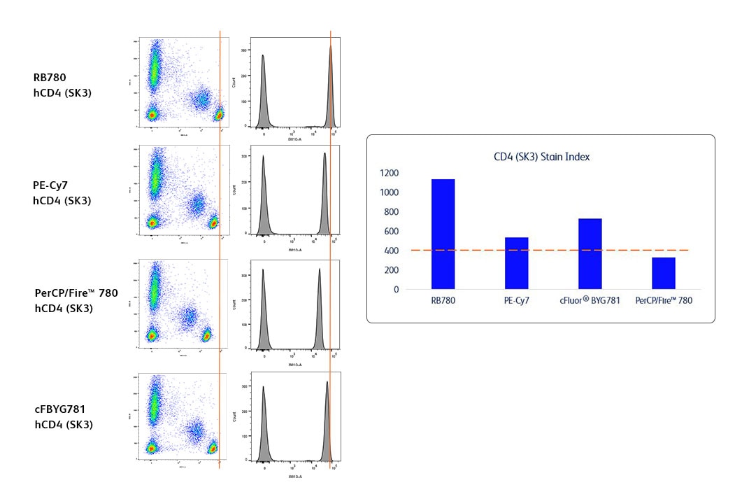
Human whole blood was stained with BD Horizon™ RB780 Mouse Anti-Human CD4 SK3 (Cat. No. 568675), BD Pharmingen™ PE-Cy7 Mouse Anti-Human CD4 SK3 (Cat. No. 557852), BioLegend PerCP/Fire™ 780 Mouse Anti-Human CD4 SK3 or Cytek cFluor® BYG781 Mouse Anti-Human CD4 SK3 Reagent. Erythrocytes were lysed with BD Pharm Lyse™ Lysing Buffer (Cat. No. 555899). The bivariate pseudocolor plots showing the expression of CD4 versus side light-scatter (SSC-A) signals were derived from gated events with the forward and side light-scatter characteristics of viable leukocytes. Histograms were derived from gated events based on light scattering characteristics for lymphocytes. Flow cytometry was performed conventionally using a BD FACSymphony™ A5 SE Cell Analyzer System. Data were generated from the Blue 810 detector using FlowJo™ Software.
While all the fluorochromes used in this experiment provided good population resolution of the CD4 positive cells, there were some differences in terms of the stain index values. We, therefore, ranked RB780, cFBYG781 and PE-Cy7 as the winners, with PerCP/Fire™ 780 as the runner up.
| Game | Spillover | |||||
| Competitors | BD Horizon RealBlue™ 705 (RB705) | BD Horizon Brilliant™ Blue 700 (BB700) | Bio-Rad StarBright™ Blue 700 (SBB700) | BD Pharmingen™ PerCP-Cy5.5 | Thermo Fisher PerCP-eFluor™ 710 | Thermo Fisher NovaFluor™ Blue 690 (NFB690) |
Rationale
Spillover was evaluated and ranked based on the analysis of a given fluorochrome’s full emission profile across five lasers. Fluorochromes with less cross-laser excitation are cleaner and will have lower impact on other fluorochromes.
We evaluated several fluorochromes primarily excited by the blue laser that have peak emission around 700 nm:
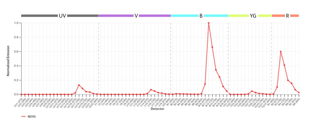
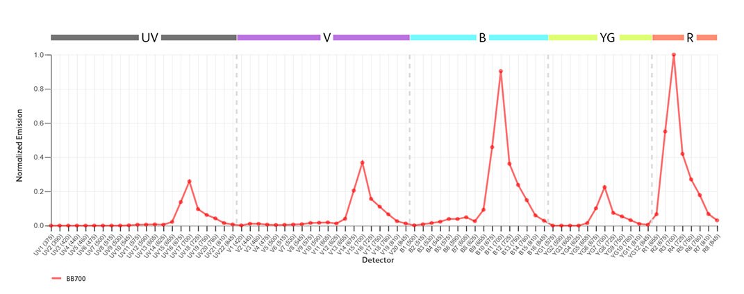
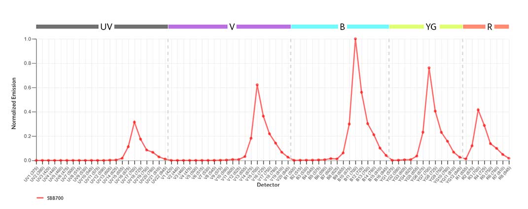
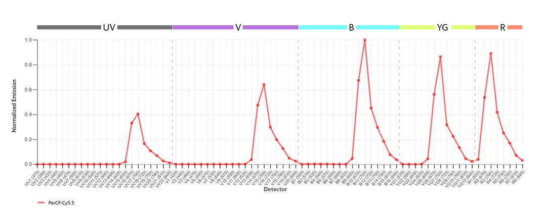
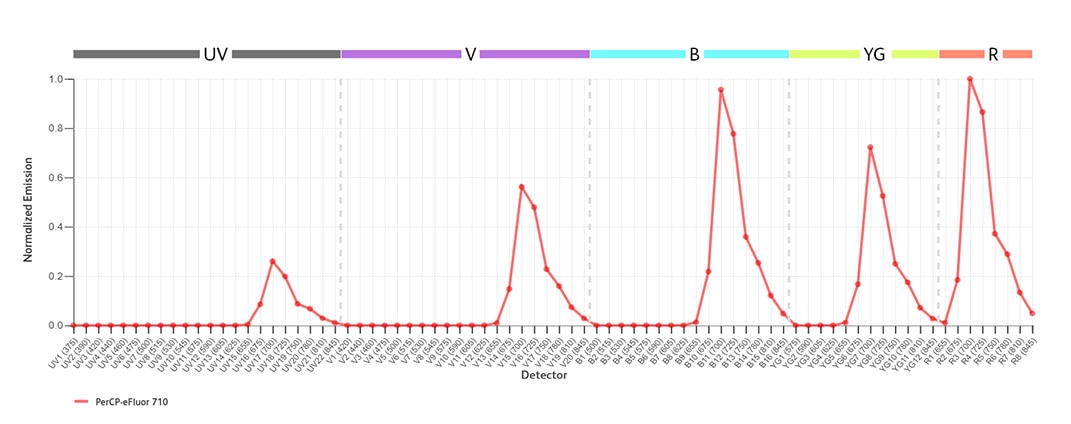
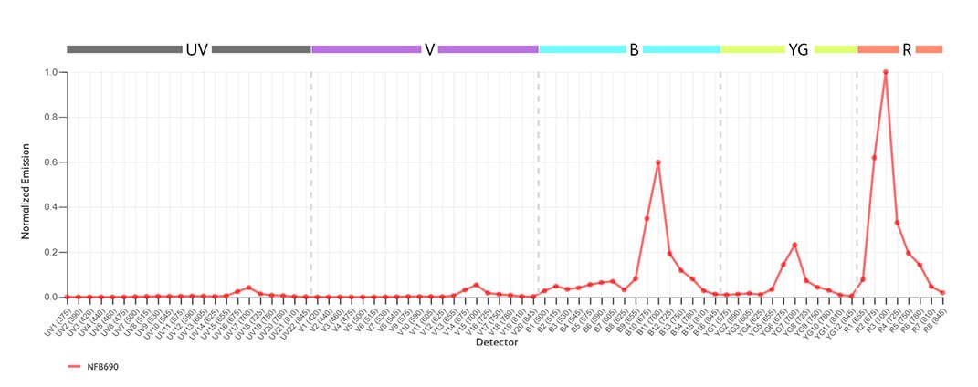
Freshly isolated human PBMCs were stained with either BD Horizon™ RB705 Mouse Anti-Human CD4 SK3 (Cat. No. 570221), BD Pharmingen™ PerCP-Cy5.5 Mouse Anti-Human CD4 SK3 (Cat. No. 566923), BD Horizon™ BB700 Mouse Anti-Human CD4 SK3 (Cat. No. 566392), Thermo Fisher PerCP-eFluor™ 710 Mouse Anti-Human CD4 SK3, Bio-Rad StarBright™ Blue 700 Mouse Anti-Human CD4 RPA-T4, or Thermo Fisher NovaFluor™ Blue 690 Anti-Human CD4 SK3 Reagent. Spectral profiles are generated from the positive population with the negative subtracted and are normalized to peak detector. Spillover percentages are dependent on the configuration of an instrument. The rankings were based on analysis on three instruments (BD FACSDiscover™ S8 Cell Sorter, BD FACSymphony™ A5 SE Cell Analyzer and Cytek Aurora). For the data shown, flow cytometry and data analysis were performed using a BD FACSDiscover™ S8 Cell Sorter and FlowJo™ Software.
Using normalized emission profiles, winners were determined based on the number of lasers with an additional peak of signal greater than 15% of the main peak signal. RB705 and NFB690 had the least amount of emission into other channels and were therefore declared the winners!
| Game | Photostability | ||
| Competitors | BD Horizon RealYellow™ 703 (RY703) | Bio-Rad StarBright™ Yellow 720 (SBY720) | Thermo Fisher NovaFluor™ Yellow 700 (NFY700) |
Rationale
We evaluated the impact of light on of fluorochromes that are primarily excited by the yellow-green laser with peak emission around 700 nm.
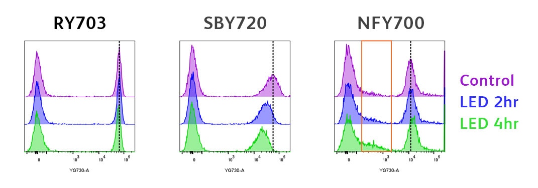
Human whole blood was stained with BD Horizon™ RY703 Mouse Anti-Human CD4 SK3 (Cat. No. 571427), Bio-Rad StarBright™ Yellow 720 Mouse Anti-Human CD4 RPA-T4 or Thermo Fisher NovaFluor™ Y700 Mouse Anti-Human CD4 SK3 Reagent. Erythrocytes were lysed with BD Pharm Lyse™ Lysing Buffer (Cat. No. 555899). After staining was completed, cells were either kept in the dark at room temperature (purple histogram) or exposed to 200 lux of LED light at room temperature for 2 (blue histogram) or 4 hours (green histogram). Histograms were derived from gated events based on light scattering characteristics for lymphocytes. Flow cytometry and data analysis were performed using a BD FACSymphony™ A5 SE Cell Analyzer System and FlowJo™ Software.
Note that in the histograms above, both MFI loss and an increase in background with light exposure can be observed. Both changes result in lower stain index and resolution.
 Change in spillover was evaluated following light exposure. This graph shows the absolute change in percent spillover into each channel after 2 hours of LED light exposure when compared to control samples kept in the dark.
T SBY720 spillover was the most impacted by light exposure and the fluorochrome also exhibited the greatest loss in resolution
Our winner for this Fluorochrome Faceoff was RY703 because this fluorochrome exhibited the least amount of spillover change and MFI loss across the dyes tested in this game.
| Game | Intracellular Staining | ||
| Competitors | BD Horizon RealBlue™ 613 (RB613) | BD Horizon™ PE-CF594 | BioLegend PE/Dazzle™ 594 |
Rationale
Here, we paired fluorochromes that are excited off the blue laser and emit at around 610 nm. We selected several intracellular markers to evaluate and stained them with three different fluorochromes. Each dye provided good population resolution and the expected staining pattern.
Note that there were clone differences between the dyes.
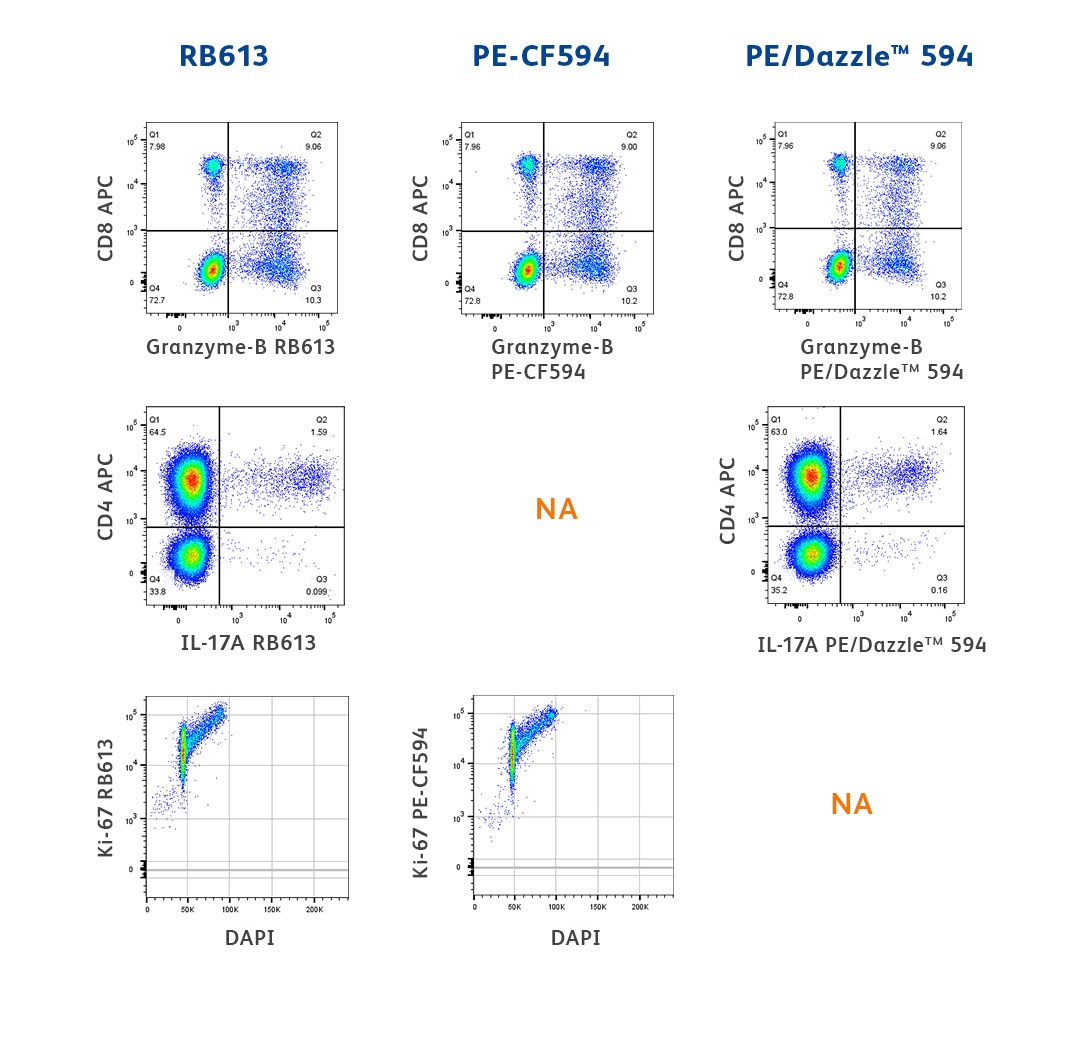
Top: Two-color flow cytometric analysis of Granzyme B expression in human peripheral blood lymphocytes. Human whole blood was fixed with BD Phosflow™ Lyse/Fix Buffer (Cat. No. 558049). The cells were then washed and stained in BD Perm/Wash™ Buffer (Cat. No. 554723) with APC Mouse Anti-Human CD8, clone RPA-T8 (Cat. No. 561953) and BD Horizon™ RB613 Mouse Anti-Human Granzyme B, clone GB11 (Cat. No. 571117), BD Horizon™ PE-CF594 Mouse Anti-Human Granzyme B, clone GB11 (Cat. No. 562462) or BioLegend PE/Dazzle™ 594 Mouse Anti-Human Granzyme B, clone QA18A28 Reagent. The bivariate pseudocolor density plots showing the correlated expression of Granzyme B versus CD8 were derived from gated events with the forward and side light-scatter characteristics of intact lymphocytes.
Middle: Two-color flow cytometric analysis of IL-17A expression in stimulated human peripheral blood lymphocytes. Human peripheral blood mononuclear cells were stimulated for 5 hours with Phorbol 12-Myristate 13-Acetate (Sigma P-8139; 50 ng/ml final concentration) and Ionomycin (Sigma I-0634; 1 µg/ml final concentration) in the presence of BD GolgiStop™ Protein Transport Inhibitor (containing Monensin) (Cat. No. 554724). The cells were harvested, washed with BD Pharmingen™ Stain Buffer (FBS) (Cat. No. 554656) and fixed with BD Cytofix™ Fixation Buffer (Cat. No. 554655). The cells were washed then permeabilized and stained in BD Perm/Wash™ Buffer (Cat. No. 554723) with APC Mouse Anti-Human CD4 SK3 antibody (Cat. No. 555349) and either BD Horizon™ RB613 Mouse Anti-Human IL-17A, clone N49-653 (Cat. No. 571130) or BioLegend PE/Dazzle™ 594 Mouse Anti-Human IL-17A, clone BL168 Reagent. The bivariate pseudocolor density plots showing the correlated expression of IL-17A versus CD4 were derived from gated events with the forward and side light-scatter characteristics of intact lymphocytes. NA indicates that the specificity/format combination was not available to us at the time of testing.
Bottom: Two-color flow cytometric analysis of Ki-67 expression by proliferating human MOLT-4 cells. Proliferating cells from the human MOLT-4 (T lymphoblastic leukemia, ATCC® CRL-1582™) cell line were permeabilized and fixed with 70% ice-cold ethanol. The cells were washed twice with BD Pharmingen™ Stain Buffer (Cat. No. 554656), stained with BD Horizon™ RB613 Mouse Anti-Ki-67, clone B56 (Cat No. 571126), BD Horizon™ PE-CF594 Mouse Anti-Ki-67, clone B56 (Cat. No. 567120) or BioLegend PE/Dazzle™ 594 Mouse Anti-Ki-67, clone 11F6 Reagent and counterstained with BD Pharmingen™ DAPI Solution (Cat. No. 564907) to stain DNA. Bivariate pseudocolor density plots showing the correlated expression of DAPI staining versus Ki-67 expression were derived from gated events with the forward and side light-scatter characteristics of MOLT-4 cells. NA indicates that the specificity/format combination was not available to us at the time of testing.
Flow cytometry and data analysis were performed using a BD FACSymphony™ A5 SE Cell Analyzer System and FlowJo™ Software.
Because all three of the dyes tested performed well for intracellular staining, we classified them all as winners! Please note that as of July 26, 2024, StarBright™ and NovaFluor™ Dye-conjugated antibodies are only available against cell surface markers and are not currently conjugated to intracellular markers. They were, therefore, not included in this Fluorochrome Faceoff.
| Game | Buffer Compatibility | ||||
| Competitors | BD Horizon RealYellow™ 610 (RY610) | BD Horizon™ PE-CF594 | Thermo Fisher NovaFluor™ Yellow 610 (NFY610) | Bio-Rad StarBright™ Yellow 605 (SBY605) | Thermo Fisher PE-eFluor™ 610 |
Rationale
Here we chose an example using a common fixation and permeabilization protocol. Staining of several fluorochromes excited by the yellow-green laser with peak emission around 610 nm was evaluated in cells treated with fixative and methanol-based permeabilization buffer after surface staining (blue line). Untreated cells were used as a reference (red line).
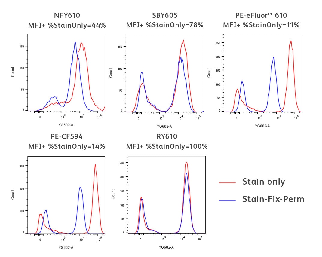
Freshly isolated human PBMCs were stained for 30 minutes with either BD Horizon™ RY610 Mouse Anti-Human CD3 UCHT1 (Cat. No. 571134), BD Horizon™ PE-CF594 Mouse Anti-Human CD3 UCHT1 (Cat. No. 562280), Thermo Fisher NovaFluor™ Yellow 610 Mouse Anti-Human CD3 UCHT1, Bio-Rad StarBright™ Yellow 605 Mouse Anti-Human CD3 UCHT1 or Thermo Fisher PE-eFluor™ 610 Mouse Anti-Human CD3 UCHT1 Reagent. The stained PBMCs were then washed twice with BD Pharmingen™ Stain Buffer (Cat. No. 554656) (red histograms). For the treated condition (blue histograms), after washing twice with BD Pharmingen™ Stain Buffer, cells were fixed using BD Cytofix™ Fixation Buffer (Cat. No. 554655) and permeabilized with BD Phosflow™ Perm Buffer III (Cat. No. 558050) for 30 minutes on ice. Cells were then washed twice with BD Pharmingen™ Stain Buffer. Histograms were derived from gated events based on light scattering characteristics for lymphocytes. Flow cytometry and data analysis were performed using a BD FACSymphony™ A5 SE Cell Analyzer System and FlowJo™ Software.
You can tell if the biological resolution of your data is impacted by the buffer you are using by comparing the staining profiles of the untreated (red) and fix-perm treated samples (blue). As you can see in the data above, there was varying impact to the fluorochromes used in this experiment, from little-to-no impact for RY610 and StarBright™ Yellow 605 to a significant reduction in signal for PE-CF594 and PE-eFluor™ 610. Therefore RY610 and StarBright™ Yellow 605 were declared as winners.
| Game | Monocyte Background | |||
| Competitors | BD Horizon RealYellow™ 775 (RY775) | BD Pharmingen™ PE-Cy7 | Bio-Rad StarBright™ Yellow 800 (SBY800) | BioLegend PE/Fire™ 810 |
Rationale
Here, we evaluated dyes excited by the yellow-green laser with peak emission around 780 nm on several clones.
As expected, PE-Cy7 demonstrated significant non-specific monocyte binding, irrespective of the antibody clone it was conjugated to. Bio-Rad StarBright™ Yellow 800 and BioLegend PE/Fire™ 810 also demonstrated varying degrees of nonspecific binding to the monocyte population.
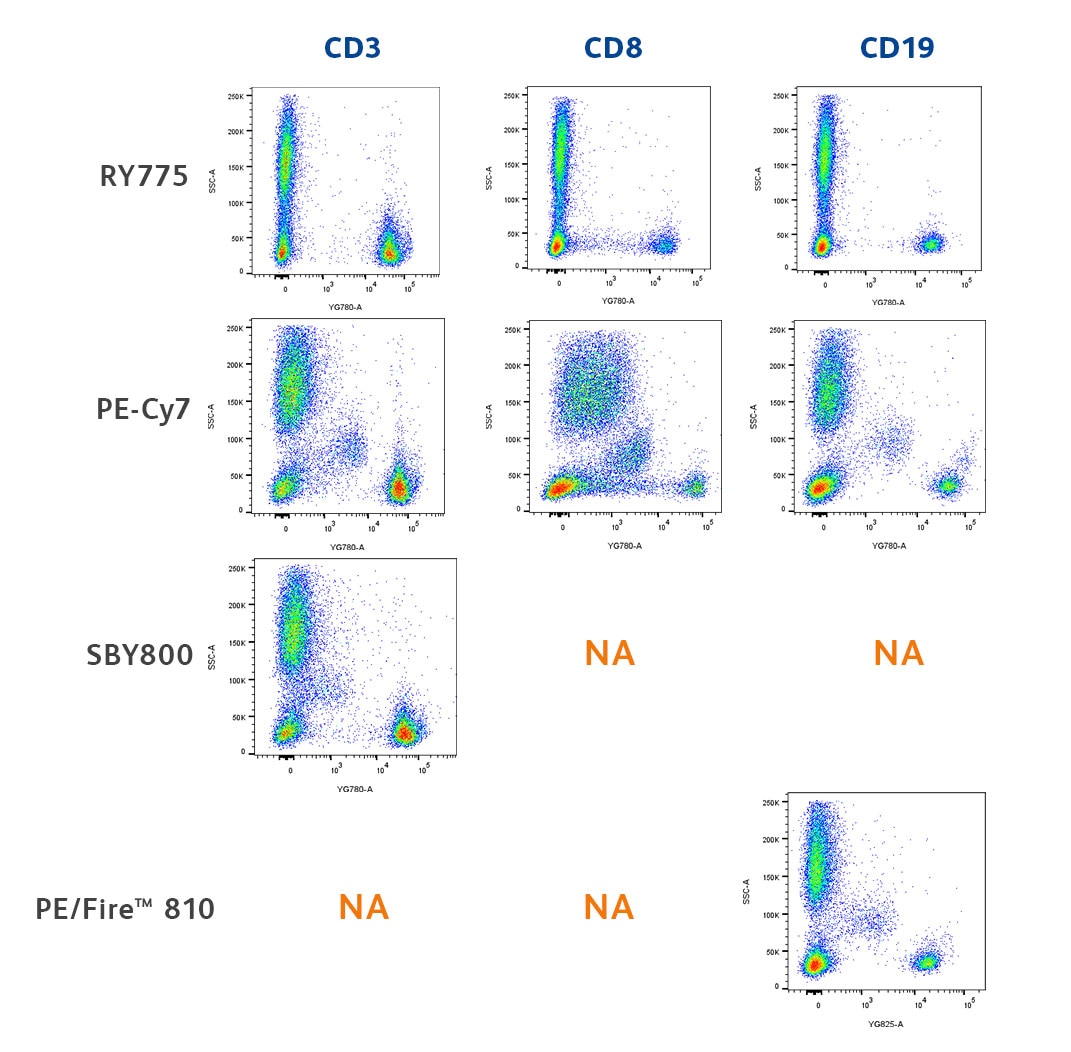
Human whole blood was stained with BD Horizon™ RY775 Mouse Anti-Human CD3 UCHT1 (Cat. No. 571376), BD Horizon™ RY775 Mouse Anti-Human CD8 RPA-T8 (Cat. No. 571380), BD Horizon™ RY775 Mouse Anti-Human CD19 SJ25C1 (Cat. No. 571374), BD Pharmingen™ PE-Cy7 Mouse Anti-Human CD3 UCHT1 (Cat. No. 563423), BD Pharmingen™ PE-Cy7 Mouse Anti-Human CD8 RPA-T8 (Cat. No. 557746), BD Pharmingen™ PE-Cy7 Mouse Anti-Human CD19 SJ25C1 (Cat. No. 557835), Bio-Rad StarBright™ Yellow 800 Mouse Anti-Human CD3 UCHT1 or BioLegend PE/Fire™ 810 Mouse Anti-Human CD19 SJ25C1 Reagent. Erythrocytes were lysed with BD Pharm Lyse™ Lysing Buffer (Cat. No. 555899). The bivariate pseudocolor plots showing the expression of CD3, CD8 and CD19 versus side light-scatter (SSC-A) signals were derived from gated events with the forward and side light-scatter characteristics of viable leukocytes. Flow cytometry and data analysis were performed using a BD FACSymphony™ A5 SE Cell Analyzer System and FlowJo™ Software. N/A means the corresponding clone/specificity was unavailable conjugated to that fluorochrome at the time of our testing.
BD Horizon RealYellow™ 775 was the only fluorochrome that did not demonstrate nonspecific background binding to monocytes and was declared the winner of this Fluorochrome Faceoff!
Conclusion
Not all dyes are created equal
BD Horizon RealYellow™ and RealBlue™ Dyes achieved a winning ranking in every faceoff, whereas other fluorochrome options had sporadic successes, winning in one or two instances but not even placing in others. Learn more about these fluorochromes and incorporate them into your panel today.