-
Reagents
- Flow Cytometry Reagents
-
Western Blotting and Molecular Reagents
- Immunoassay Reagents
-
Single-Cell Multiomics Reagents
- BD® OMICS-Guard Sample Preservation Buffer
- BD® AbSeq Assay
- BD® Single-Cell Multiplexing Kit
- BD Rhapsody™ ATAC-Seq Assays
- BD Rhapsody™ Whole Transcriptome Analysis (WTA) Amplification Kit
- BD Rhapsody™ TCR/BCR Next Multiomic Assays
- BD Rhapsody™ Targeted mRNA Kits
- BD Rhapsody™ Accessory Kits
- BD® OMICS-One Protein Panels
-
Functional Assays
-
Microscopy and Imaging Reagents
-
Cell Preparation and Separation Reagents
-
- BD® OMICS-Guard Sample Preservation Buffer
- BD® AbSeq Assay
- BD® Single-Cell Multiplexing Kit
- BD Rhapsody™ ATAC-Seq Assays
- BD Rhapsody™ Whole Transcriptome Analysis (WTA) Amplification Kit
- BD Rhapsody™ TCR/BCR Next Multiomic Assays
- BD Rhapsody™ Targeted mRNA Kits
- BD Rhapsody™ Accessory Kits
- BD® OMICS-One Protein Panels
- Italy (English)
-
Change country/language
Old Browser
This page has been recently translated and is available in French now.
Looks like you're visiting us from United States.
Would you like to stay on the current country site or be switched to your country?
BD® OMICS-One T-cell Protein Panel


Regulatory Status Legend
Any use of products other than the permitted use without the express written authorization of Becton, Dickinson and Company is strictly prohibited.
Product Details
Description
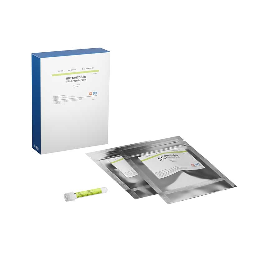
The BD® OMICS-One T-Cell Protein Panel consists of 30 different specificities against major T-cell markers in a single tube. Designed and optimized to work on the BD Rhapsody™ System, the T-Cell Protein Panel is tested to work seamlessly alongside the BD Rhapsody™ Whole Transcriptome Analysis (WTA) Assay, Targeted mRNA Assay, BD® Single-Cell Multiplexing Kit (SMK), BD® Intracellular CITE-seq (IC-AbSeq) Assay, and BD Rhapsody™ TCR/BCR Next Multiomic Assay for humans. The individual antibodies were each conjugated to an oligonucleotide that contains a specific antibody barcode sequence flanked by a polyA tail on the 3' end and a common PCR handle (PCR primer binding site) on the 5' end. To allow for sequencing error correction and unique mapping, all AbSeq barcode sequences were generated in silico with minimal sequence similarity to the human genomes, have low predicted secondary structure, and have high Hamming distance within the BD antibody-oligo portfolio. The polyA tail of the oligonucleotide allows the barcode sequence to be captured by the BD Rhapsody™ Enhanced Cell Capture Beads. The 5' PCR handle allows for efficient sequencing library generation for various sequencing platforms. Each individual antibody exists at an optimal concentration within the 30-plex to enable superior target and population resolution.
The T-Cell Protein Panel is designed with SMART technology. SMART technology helps lower sequencing cost while increasing data resolution by attenuating antibodies that target high-expressing primary markers and by allowing re-allocation of sequencing reads to markers expressed at lower levels. With SMART technology, markers low in expression can be quantified without having to do deeper sequencing and incurring high sequencing cost. The two specificities attenuated in the T-Cell Protein Panel are CD4 and CD44.
Preparation And Storage
Recommended Assay Procedures
This reagent is provided lyophilized in a pre-titrated format.
1. Remove the BD® OMICS-One T-Cell Protein Panel tube from the foil bag and bring up to room temperature for 5 minutes.
2. Make sure the pellet is located at the bottom of the tube. If not, briefly centrifuge to collect the contents at the tube bottom.
3. Add 35 µL of nuclease-free water to the bottom of the tube and allow antibodies to reconstitute for 5 minutes at room temperature.
4. Transfer the reconstituted antibodies on ice until the cells are ready for staining.
Note: Reconstitute antibodies immediately before cell staining. Prolonged incubation of reconstituted antibody might increase the
non-specific background.
5. For BD® AbSeq Ab-Oligo drop-in of 60 plex or lower, prepare the BD® AbSeq labeling MasterMix in a 1.5-mL LoBind tube on ice.
Note: For drop-in with more than 60 plex, reach out to technical support for calculation.
For sequential labeling with Sample Tags or no Sample Tags, prepare BD® AbSeq labeling MasterMix for drop-ins as follows:
____________________________________________________________________________________________
Component 1 sample (µL) 1 sample + 2 samples +
30% overage (µL) 30% overage (µL)
Per BD® AbSeq Ab-Oligo 2.0 2.6 5.2
Total of BD® AbSeq Ab-Oligo 2.0 × N* 2.6 × N 5.2 × N
FBS† (catalog number 554656) 140 – (2.0 x N) 182 – (2.6 x N) 364 – (5.2 x N)
Total 140 182 364
For co-labeling with Sample Tags, prepare BD® AbSeq labeling MasterMix for drop-ins as follows:
____________________________________________________________________________________________
Component 1 sample (µL) 1 sample + 2 samples +
30% overage (µL) 30% overage (µL)
Per BD® AbSeq Ab-Oligo 2.0 2.6 5.2
Total of BD® AbSeq Ab-Oligo 2.0 × N* 2.6 × N 5.2 × N
FBS† (catalog number 554656) 120 – (2.0 × N) 156 – (2.6 × N) 312 – (5.2 × N)
Total 120 156 312
* N = number of drop-in antibodies. N = 0 if there are no drop-in antibodies.
† FBS = BD Pharmingen™ Stain Buffer.
6. Pipet-mix the BD® AbSeq labeling MasterMix for drop-ins. Briefly centrifuge to collect the contents at the bottom, and place back on ice.
7. For sequential labeling with Sample Tags or no Sample Tags, for each sample, add 140 µL BD® AbSeq labeling MasterMix of drop-ins to
the tube containing 35 µL reconstituted T-Cell Protein Panel solution to make a total volume of 175 µL.
For co-labeling with Sample Tags, for each sample, add 120 µL BD® AbSeq labeling MasterMix of drop-ins and 20 µL Sample Tag to the
tube containing 35 µL reconstituted T-Cell Protein Panel solution to make a total volume of 175 µL.
8. Pipet-mix the mixture, briefly centrifuge to collect the contents at the tube bottom, and place back on ice.
9. Centrifuge cells at 400 × g for 5 minutes. If Fc Block is used, proceed to step 10. Otherwise, skip to step 11.
10. (Optional) For samples containing myeloid and B lymphocytes, BD Biosciences recommends blocking nonspecific Fc Receptor–mediated
false-positive signals with Human BD Fc Block (Cat. No. 564220).
a. To perform blocking, pipet the Fc Block MasterMix into a new 1.5-mL LoBind tube on ice:
_________________________________________________________________________________
Component 1 sample (µL)* 1 sample + 20% overage (µL)
FBS† (catalog number 554656) 20.0 24.0
Fc Block‡ (catalog number 564220) 5.0 6.0
Total 25.0 30.0
* Sufficient for up to 1 million cells. To block more cells, adjust the volume.
† FBS = BD Pharmingen™ Stain Buffer.
‡ Fc Block = BD Pharmingen™ Human BD Fc Block.
b. Pipet-mix the Fc Block MasterMix and briefly centrifuge. Place on ice.
c. Remove the supernatant from the cells without disturbing the pellet.
d. Resuspend the cells in 25 µL of Fc Block MasterMix.
e. Incubate the cells at room temperature (15°C to 25°C) for 10 minutes.
f. Add 175 µL of BD® AbSeq labeling MasterMix from Step 8 into the cell suspension. Pipet-mix and proceed to Step 12.
11. Remove the supernatant from the cells without disturbing the pellet. Add 25 µL Stain Buffer (FBS) to the 175 µL of BD® AbSeq labeling
MasterMix from Step 8 to make a total volume of 200 µL. Resuspend the cell pellet in 200 µL total volume. Pipet-mix.
12. Transfer the cells with BD® AbSeq labeling MasterMix into a new 5-mL polystyrene Falcon tube.
13. Stain the cells on ice for 30 minutes.
14. Add 3–4 mL Stain Buffer (FBS) to labelled cells and pipet-mix.
15. Centrifuge at 400 x g for 5 minutes.
16. Uncap the tube and invert to decant supernatant into biohazardous waste. Keep the tube inverted and gently blot on a lint-free wiper to
remove residual supernatant from tube rim.
17. Repeat steps 14–16 twice more for a total of three washes.
18. Resuspend the final washed cell pellet in 620 µL cold Sample Buffer from the BD Rhapsody™ Enhanced Cartridge Reagent V3
(catalog number 667052) and proceed to single cell capture with on-cartridge washing described in substeps a–c. See BD Rhapsody™ HT
Single-Cell Analysis System Single-Cell Capture and cDNA Synthesis Protocol (Doc ID 23-24252) or BD Rhapsody™ HT Xpress System
Single-Cell Capture and cDNA Synthesis Protocol (Doc ID 23-24253) for additional details.
Note: Perform on-cartridge washing after cell settling (8 minute incubation) as follows:
a. At the protocol section of "Loading cells in BD Rhapsody™ 8-Lane Cartridge", after cell load, incubate the cartridge in the dark
at room temperature for 8 minutes.
b. Place the cartridge on the BD Rhapsody™ HT Xpress and perform the following operations:
_____________________________________________________________
Material to load Volume (µL) 1 lane Pipette Mode
Air 380 Prime/Wash
Cold Sample Buffer 380 Prime/Wash
Air 380 Prime/Wash
Cold Sample Buffer 380 Prime/Wash
c. (Optional) Perform the scanner step: Cell Load Scan, if using BD Rhapsody™ HT Single-Cell Analysis System Single-Cell
Capture and cDNA Synthesis Protocol (Doc ID 23-24252). No need for 8-minute delay before scanning.
Warning: All biological specimens and materials are considered biohazardous. Handle as if capable of transmitting infection and dispose using proper precautions in accordance with federal, state, and local regulations. Never pipette by mouth. Wear suitable protective clothing, eyewear, and gloves.
List of all 30 Human AbSeq specificities included in the BD® OMICS-One T-Cell panel:
_____________________________________________________________________________
Specificity Clone Oligo ID BD® AbSeq Barcode Sequence
CD103 Ber-ACT8 AHS0001 AAATAGTATCGAGCGTAGTTAAGTTGCGTAGCCGTT
CD137 4B4-1 AHS0003 TGACAAGCAACGAGCGATACGAAAGGCGAAATTAGT
CD45RA HI100 AHS0009 AAGCGATTGCGAAGGGTTAGTCAGTACGTTATGTTG
CD69 FN50 AHS0010 CAATAACGGGTCATAGTAAGTCGCGAGTAAGAGGGC
CD278 DX29 AHS0012 ATAGTCCGCCGTAATCGTTGTGTCGCTGAAAGGGTT
CD134 (OX40) ACT35 AHS0013 GGTGTTGGTAAGACGGACGGAGTAGATATTCGAGGT
CD279 (PD-1) EH12.1 AHS0014 ATGGTAGTATCACGACGTAGTAGGGTAATTGGCAGT
CD366 (TIM-3) 7D3 AHS0016 TAGGTAGTAGTCCCGTATATCCGATCCGTGTTGTTT
CD223 (Lag3) T47-530 AHS0018 CGGCATGAATTAGGCGAGACTTAGTATACGAGCTGG
CD95 (Fas) DX2 AHS0023 GGCCCGTTAGAGTTGGTATCCGTATGAAGGTTAGCT
CD25 2A3 AHS0026 AGTTGTATGGGTTAGCCGAGAGTAGTGCGTATGATT
CD127 HIL-7R-M21 AHS0028 AGTTATTAGGCTCGTAGGTATGTTTAGGTTATCGCG
CD183 1C6/CXCR3 AHS0031 AAAGTGTTGGCGTTATGTGTTCGTTAGCGGTGTGGG
CD4 SK3 AHS0032 TCGGTGTTATGAGTAGGTCGTCGTGCGGTTTGATGT
CD196 (CCR6) 11A9 AHS0034 ACGTGTTATGGTGTTGTTCGAATTGTGGTAGTCAGT
CD45RO UCHL1 AHS0036 TGAGAGGTTATTGGGCGTATGACTTCGGTGATTGTG
CD194 (CCR4) 1G1 AHS0038 AATATTAGTGGGTCCTCGCGTTGGCCGGTTGTTAGT
CD62L DREG-56 AHS0049 ATGGTAAATATGGGCGAATGCGGGTTGTGCTAAAGT
CD272 J168-540 AHS0052 GTAGGTTGATAGTCGGCGATAGTGCGGTTGAAAGCT
CD154 TRAP1 AHS0077 TAAGAGGTAAGTGCATTCGGGTATAGGCGTGATTTG
CD357 (GITR) V27-580 AHS0104 TCTGTGTGTCGGGTTGAATCGTAGTGAGTTAGCGTG
CD28 L293 AHS0138 TTGTTGAGGATACGATGAAGCGGTTTAAGGGTGTGG
TCRgd 11F2 AHS0142 CTCGTGGGTTAGGCTTGATCGTAGTTATGTATGGTT
CD44 L178 AHS0167 GTGATTGATTAGGACAGTTCGTTGCTTAGTAGTGGG
TCR Va24-Ja18 6B11 AHS0175 TTCTGGTTCGGTTGAGCTACTAATTTCGTTGGATGG
CD161 (KLRB1) HP-3G10 AHS0205 TTTAGGACGATTAGTTGTGCGGCATAGGAGGTGTTC
CD8 SK1 AHS0228 AGGACATAGAGTAGGACGAGGTAGGCTTAAATTGCT
CD3 UCHT1 AHS0231 AGCTAGGTGTTATCGGCAAGTTGTACGGTGAAGTCG
CD197 (CCR7) 2-L1-A AHS0273 AATGTGTGATCGGCAAAGGGTTCTCGGGTTAATATG
TIGIT tgMab-2 AHS0411 AGAGGGTTTAGTCAAGGTCGTGCGTATAGTTCAGGT
Product Notices
- This reagent is provided lyophilized in a pre-titrated format.
- Please refer to https://www.bdbiosciences.com/en-us/resources/protocols/single-cell-multiomics for technical protocols.
- The production process underwent stringent testing and validation to assure that it generates a high-quality conjugate with consistent performance and specific binding activity. However, verification testing has not been performed on all conjugate lots.
- Go to https://abseq-ref-gen.genomics.bd.com/to access AbSeq reference files in FASTA format for bioinformatics analyses.
- Caution: Sodium azide yields highly toxic hydrazoic acid under acidic conditions. Dilute azide compounds in running water before discarding to avoid accumulation of potentially explosive deposits in plumbing. Follow state and local guidelines when disposing of hazardous waste.
- Source of all serum proteins is from USDA inspected abattoirs located in the United States.
- For U.S. patents that may apply, see bd.com/patents.
- Please refer to http://regdocs.bd.com to access safety data sheets (SDS).
Please refer to Support Documents for Quality Certificates
Global - Refer to manufacturer's instructions for use and related User Manuals and Technical data sheets before using this products as described
Comparisons, where applicable, are made against older BD Technology, manual methods or are general performance claims. Comparisons are not made against non-BD technologies, unless otherwise noted.
For Research Use Only. Not for use in diagnostic or therapeutic procedures.