-
Reagents
- Flow Cytometry Reagents
-
Western Blotting and Molecular Reagents
- Immunoassay Reagents
-
Single-Cell Multiomics Reagents
- BD® OMICS-Guard Sample Preservation Buffer
- BD® AbSeq Assay
- BD® Single-Cell Multiplexing Kit
- BD Rhapsody™ ATAC-Seq Assays
- BD Rhapsody™ Whole Transcriptome Analysis (WTA) Amplification Kit
- BD Rhapsody™ TCR/BCR Next Multiomic Assays
- BD Rhapsody™ Targeted mRNA Kits
- BD Rhapsody™ Accessory Kits
- BD® OMICS-One Protein Panels
-
Functional Assays
-
Microscopy and Imaging Reagents
-
Cell Preparation and Separation Reagents
-
- BD® OMICS-Guard Sample Preservation Buffer
- BD® AbSeq Assay
- BD® Single-Cell Multiplexing Kit
- BD Rhapsody™ ATAC-Seq Assays
- BD Rhapsody™ Whole Transcriptome Analysis (WTA) Amplification Kit
- BD Rhapsody™ TCR/BCR Next Multiomic Assays
- BD Rhapsody™ Targeted mRNA Kits
- BD Rhapsody™ Accessory Kits
- BD® OMICS-One Protein Panels
- Italy (English)
-
Change location/language
Old Browser
This page has been recently translated and is available in French now.
Looks like you're visiting us from {countryName}.
Would you like to stay on the current location site or be switched to your location?
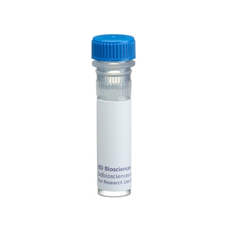
BD Transduction Laboratories™ Purified Mouse Anti-IAK1
Clone 4/IAK1 (RUO)


Western blot analysis of IAK1 on a Jurkat cell lysate (Human T-cell leukemia; ATCC TIB-152). Lane 1: 1:250, lane 2: 1:500, lane 3: 1:1000 dilution of the mouse anti-IAK1 antibody.

Immunofluorescence staining of human endothelial cells.


Regulatory Status Legend
Any use of products other than the permitted use without the express written authorization of Becton, Dickinson and Company is strictly prohibited.
Preparation And Storage
Recommended Assay Procedures
Western blot: Please refer to http://www.bdbiosciences.com/pharmingen/protocols/Western_Blotting.shtml
Product Notices
- Since applications vary, each investigator should titrate the reagent to obtain optimal results.
- Please refer to www.bdbiosciences.com/us/s/resources for technical protocols.
- Caution: Sodium azide yields highly toxic hydrazoic acid under acidic conditions. Dilute azide compounds in running water before discarding to avoid accumulation of potentially explosive deposits in plumbing.
- Source of all serum proteins is from USDA inspected abattoirs located in the United States.
Companion Products


Cell division is a tightly regulated process that ensures the segregation of chromosomes into daughter cells. Essential to this regulation is the modification of cell cycle components by reversible phosphorylation. Ipl1 and aurora are two related kinases isolated from S.cerevisiae and Drosophila, respectively. Inactivation of these kinases results in abnormal chromosome segregation and disruption of the centrosome. A structurally and functionally similar kinase, IAK1 (Ipl1- and Aurora-related kinase 1), is a regulator of mammalian chromosome segregation. Although IAK1 may be present in the cytoplasm, it is detected on the centrosome following duplication and also associates with the spindle microtubules from metaphase through cell division. Expression of IAK1 is stringently regulated during the cell cycle. Both mRNA and protein are initially expressed in S-phase, are elevated during M-phase, and are undetectable following completion of mitosis. Increasing evidence suggests that IAK1 belongs to a novel subfamily of the ser/thr kinase superfamily. Although mutational analysis of IAK1 will directly determine its function, it appears to be a key player in the control of cell division.
Development References (4)
-
Gopalan G, Chan CS, Donovan PJ. A novel mammalian, mitotic spindle-associated kinase is related to yeast and fly chromosome segregation regulators.. J Cell Biol. 1997; 138(3):643-56. (Biology). View Reference
-
Katayama H, Ota T, Jisaki F. Mitotic kinase expression and colorectal cancer progression. J Natl Cancer Inst. 1999; 91(13):1160-1162. (Biology: Western blot). View Reference
-
Kiat LS, Hui KM, Gopalan G. Aurora-A kinase interacting protein (AIP), a novel negative regulator of human Aurora-A kinase. J Biol Chem. 2002; 277(47):4558-45565. (Biology: Western blot). View Reference
-
Sakai H, Urano T, Ookata K. MBD3 and HDAC1, two components of the NuRD complex, are localized at Aurora-A-positive centrosomes in M phase. J Biol Chem. 2002; 277(50):48714-48723. (Biology: Immunofluorescence). View Reference
Please refer to Support Documents for Quality Certificates
Global - Refer to manufacturer's instructions for use and related User Manuals and Technical data sheets before using this products as described
Comparisons, where applicable, are made against older BD Technology, manual methods or are general performance claims. Comparisons are not made against non-BD technologies, unless otherwise noted.
For Research Use Only. Not for use in diagnostic or therapeutic procedures.