BD Transduction Laboratories™ Purified Mouse Anti-VCP
Clone 18/VCP (RUO)
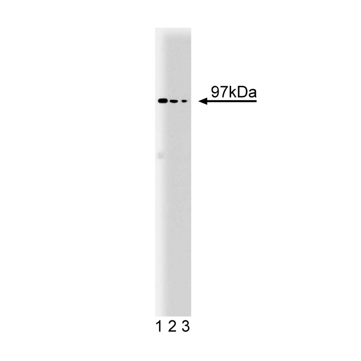
Western blot analysis of VCP on a mouse cerebrum lysate. Lane 1: 1:250, lane 2: 1:500, lane 3: 1:1000 dilution of the mouse anti-VCP antibody.
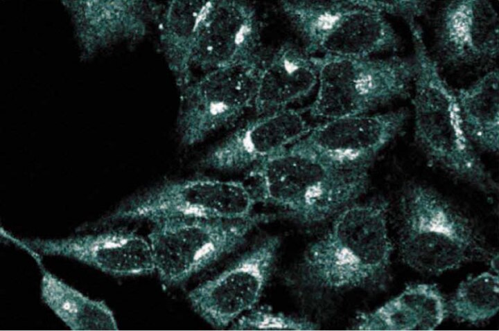
Immunofluorescence staining of HeLa cells (Human cervical epitheloid carcinoma; ATCC CCL-2.2).

Western blot analysis of VCP on a mouse cerebrum lysate. Lane 1: 1:250, lane 2: 1:500, lane 3: 1:1000 dilution of the mouse anti-VCP antibody.
Immunofluorescence staining of HeLa cells (Human cervical epitheloid carcinoma; ATCC CCL-2.2).
Preparation And Storage
Recommended Assay Procedures
Western blot: Please refer to http://www.bdbiosciences.com/pharmingen/protocols/Western_Blotting.shtml
Product Notices
- Since applications vary, each investigator should titrate the reagent to obtain optimal results.
- Please refer to www.bdbiosciences.com/us/s/resources for technical protocols.
- Source of all serum proteins is from USDA inspected abattoirs located in the United States.
- Caution: Sodium azide yields highly toxic hydrazoic acid under acidic conditions. Dilute azide compounds in running water before discarding to avoid accumulation of potentially explosive deposits in plumbing.
Companion Products


ATPases that belong to the ATPase associated with different cellular activities (AAA) family are homo-oligomeric proteins that have roles in cell cycle regulation, protein degradation, organelle biogenesis, and vesicle mediated-protein transport. Valosin containing protein (VCP) is a AAA family member that is found as a hexamer in rat liver. In vitro, VCP catalyzes the hydrolysis of ATP, but this activity is distinct from classical transport ATPases. VCP protein is found primarily in the transitional elements between the rough and smooth ER (TER), but stimulation with EGF leads to VCP translocation to the nucleus. Antibodies to VCP can perturb cell-free formation of transition vesicles from isolated TER of rat liver, and can inhibit transfer of material from ER to Golgi in a reconstituted membrane system. VCP is phosphorylated after T-cell activation, and PTPH1 dephosphorylation of VCP correlates with suppression of cell growth. In addition, the N-terminal region of VCP binds to the DNA damage repair protein, BRCA1. Thus, VCP may have roles in both ER protein transport and nuclear function, which are important for cell growth and survival.
This antibody is routinely tested by western blot analysis. Other applications were tested by BD Biosciences Pharmingen during antibody development only or reported in the literature.
Development References (4)
-
Egerton M, Ashe OR, Chen D, Druker BJ, Burgess WH, Samelson LE. VCP, the mammalian homolog of cdc48, is tyrosine phosphorylated in response to T cell antigen receptor activation. EMBO J. 1992; 11(10):3533-3540. (Biology). View Reference
-
Zhang H, Wang Q, Kajino K, Greene MI. VCP, a weak ATPase involved in multiple cellular events, interacts physically with BRCA1 in the nucleus of living cells. DNA Cell Biol. 2000; 19(5):253-263. (Biology). View Reference
-
Zhang L, Ashendel CL, Becker GW, Morre DJ. Isolation and characterization of the principal ATPase associated with transitional endoplasmic reticulum of rat liver. J Cell Biol. 1994; 127(6 Pt 2):1871-1883. (Biology). View Reference
-
Zhang SH, Liu J, Kobayashi R, Tonks NK. Identification of the cell cycle regulator VCP (p97/CDC48) as a substrate of the band 4.1-related protein-tyrosine phosphatase PTPH1. J Biol Chem. 1999; 274(25):17806-17812. (Biology). View Reference
Please refer to Support Documents for Quality Certificates
Global - Refer to manufacturer's instructions for use and related User Manuals and Technical data sheets before using this products as described
Comparisons, where applicable, are made against older BD Technology, manual methods or are general performance claims. Comparisons are not made against non-BD technologies, unless otherwise noted.
For Research Use Only. Not for use in diagnostic or therapeutic procedures.