Old Browser
This page has been recently translated and is available in French now.
Looks like you're visiting us from {countryName}.
Would you like to stay on the current country site or be switched to your country?


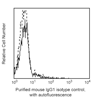
Flow cytometric analysis of CD21 expression on human peripheral blood lymphocytes. Whole blood was stained with either Purified Mouse Anti-Human CD21 (Cat. No. 555421; solid line histogram) or Purified Mouse IgG1 κ Isotype Control (Cat. No. 555746; dashed line histogram), followed by FITC Goat Anti-Mouse IgG/IgM (Cat. No. 555988). Erythrocytes were lysed with BD Pharm Lyse™ Lysing Buffer (Cat. No. 555899). Fluorescent histograms were derived from gated events with the side and forward light-scattering characteristics of viable lymphocytes. Flow cytometry was performed on a BD FACSCan™ system.


BD Pharmingen™ Purified Mouse Anti-Human CD21

Regulatory Status Legend
Any use of products other than the permitted use without the express written authorization of Becton, Dickinson and Company is strictly prohibited.
Preparation And Storage
Product Notices
- Since applications vary, each investigator should titrate the reagent to obtain optimal results.
- An isotype control should be used at the same concentration as the antibody of interest.
- Caution: Sodium azide yields highly toxic hydrazoic acid under acidic conditions. Dilute azide compounds in running water before discarding to avoid accumulation of potentially explosive deposits in plumbing.
- Sodium azide is a reversible inhibitor of oxidative metabolism; therefore, antibody preparations containing this preservative agent must not be used in cell cultures nor injected into animals. Sodium azide may be removed by washing stained cells or plate-bound antibody or dialyzing soluble antibody in sodium azide-free buffer. Since endotoxin may also affect the results of functional studies, we recommend the NA/LE (No Azide/Low Endotoxin) antibody format, if available, for in vitro and in vivo use.
- Species cross-reactivity detected in product development may not have been confirmed on every format and/or application.
- Please refer to www.bdbiosciences.com/us/s/resources for technical protocols.
Companion Products

.png?imwidth=320)
The B-ly4 monoclonal antibody specifically binds to CD21, a 145 kDa glycosylated type I integral membrane protein. CD21 is a receptor for the C3d complement fragment and for Epstein-Barr virus (EBV). CD21 is expressed on mature B cells, follicular dendritic cells, and some epithelial cells. It is also weakly expressed on the subset of mature T cells and thymocytes. CD21 plays a role in B-cell activation and proliferation. It may also play a role in modulating the function of T cells in the immune response to infections by lymphotropic viruses. Recently, CD21 was found to be part of a large complex containing CD19, CD81, and possibly other molecules.
This clone also cross-reacts with a major subset of, but not all, peripheral blood CD20+ lymphocytes of baboon, and both rhesus and cynomolgus macaque monkeys. A subset of CD3+ cells is also CD21+.
Development References (6)
-
Fearon DT. The CD19-CR2-TAPA-1 complex, CD45 and signaling by the antigen receptor of B lymphocytes. Curr Opin Immunol. 1993; 5(3):341-348. (Biology). View Reference
-
Fischer E, Delibrias C, Kazatchkine MD. Expression of CR2 (the C3dg/EBV receptor, CD21) on normal human peripheral blood T lymphocytes. J Immunol. 1991; 146(3):865-869. (Biology). View Reference
-
Knapp W. W. Knapp .. et al., ed. Leucocyte typing IV : white cell differentiation antigens. Oxford New York: Oxford University Press; 1989:1-1182.
-
Lin G-X, Yang X, Hollemweguer E, et al. Cross-reactivity of CD antibodies in eight animal species. In: Mason D. David Mason .. et al., ed. Leucocyte typing VII : white cell differentiation antigens : proceedings of the Seventh International Workshop and Conference held in Harrogate, United Kingdom. Oxford: Oxford University Press; 2002:519-523.
-
Paterson RL, Kelleher C, Amankonah TD, et al. Model of Epstein-Barr virus infection of human thymocytes: expression of viral genome and impact on cellular receptor expression in the T-lymphoblastic cell line, HPB-ALL. Blood. 1995; 85(2):456-464. (Biology). View Reference
-
Tsoukas CD, Lambris JD. Expression of EBV/C3d receptors on T cells: biological significance. Immunol Today. 1993; 14(2):56-59. (Biology). View Reference
Please refer to Support Documents for Quality Certificates
Global - Refer to manufacturer's instructions for use and related User Manuals and Technical data sheets before using this products as described
Comparisons, where applicable, are made against older BD Technology, manual methods or are general performance claims. Comparisons are not made against non-BD technologies, unless otherwise noted.
For Research Use Only. Not for use in diagnostic or therapeutic procedures.
Report a Site Issue
This form is intended to help us improve our website experience. For other support, please visit our Contact Us page.