Dear Customer, If you experience any text readability or display issues on our website, please update your web browser to the latest version
.
See More
Dear Customer, If you experience any text readability or display issues on our website, please update your web browser to the latest version
.
See MoreDear Customer, If you experience any text readability or display issues on our website, please update your web browser to the latest version
.
This page has been recently translated and is available in French now.
Looks like you're visiting us from {countryName}.
Would you like to stay on the current location site or be switched to your location?
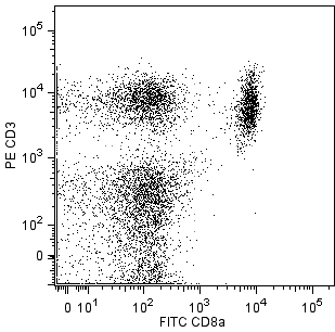
Two-color analysis of the expression of TCR Vβ3 on mouse peripheral lymphocytes. C57BL/6 lymph node cells were incubated simultaneously with PE Hamster Anti-Mouse TCR Vβ3 (Cat. No. 553209), FITC Rat Anti-Mouse CD4 (Cat. No. 553046/553047), and FITC Rat Anti-Mouse CD8a (Cat. No. 553030/553031) monoclonal antibodies. The fluorescence contour plot was derived from gated events based on the forward and side light-scattering of viable lymphocytes. Flow cytometry was performed on a BD FACScan™.
Any use of products other than the permitted use without the express written authorization of Becton, Dickinson and Company is strictly prohibited.
For flow cytometry of cell suspensions from peripheral lymphoid tissues, it is recommended that multicolor staining be performed to distinguish T lymphocytes from non-T cells.
The KJ25 antibody specifically reacts with the Vβ 3 T-cell Receptor (TCR) of strains having the a (e.g., C57BR, SJL), b (e.g., AKR, CBA/Ca, C57BL, DBA/1), and c (e.g., RIII) haplotypes of the Tcrb gene complex. Vβ 3 TCR-bearing T lymphocytes are clonally eliminated either completely or partially in mice expressing superantigens encoded by the Mtv-1 (Mls-4[a], Mls[c]), Mtv-3 (Mls[c]), Mtv-6 (Mls-3[a], Mls[c]), Mtv-13 (Mls-2[a], Mls[c]), Mtv-27, Mtv-44, and/or Mtv-MAI endogenous proviruses (e.g., A, BALB/c, CBA/J, C3H/He, DBA/2, NZB, NZW). Vβ 3 TCR-bearing T cells are activated by the superantigenic Staphylococcal Enterotoxins A and B. Activation or elimination of Vβ 3 TCR-expressing T cells by these determinants is partially dependent upon presentation by I-E. This hamster mAb to a mouse leukocyte antigen does not cross-react with rat leukocytes.
Please refer to Support Documents for Quality Certificates
Global - Refer to manufacturer's instructions for use and related User Manuals and Technical data sheets before using this products as described
Comparisons, where applicable, are made against older BD Technology, manual methods or are general performance claims. Comparisons are not made against non-BD technologies, unless otherwise noted.
For Research Use Only. Not for use in diagnostic or therapeutic procedures.