The site is undergoing maintenance on
Thursday, September 10, 7:00 pm through Friday, September 11, 3:00 am (CEST), 2026.
If you encounter problems while ordering, please contact us at BDBCustomerService@bd.com
See More
The site is undergoing maintenance on
Thursday, September 10, 7:00 pm through Friday, September 11, 3:00 am (CEST), 2026.
If you encounter problems while ordering, please contact us at BDBCustomerService@bd.com
See MoreThe site is undergoing maintenance on
Thursday, September 10, 7:00 pm through Friday, September 11, 3:00 am (CEST), 2026
.If you encounter problems while ordering, please contact us at BDBCustomerService@bd.com
This page has been recently translated and is available in French now.
Looks like you're visiting us from {countryName}.
Would you like to stay on the current location site or be switched to your location?
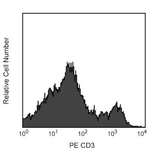
Multicolor flow cytometric analysis of CD4 expression on rat splenocytes. Splenocytes from a Lewis rat were stained with the PE-Cy™7 Mouse Anti-Rat CD4 antibody (Cat. No. 561578) in conjunction with a PE Mouse Anti-Rat CD3 antibody (Cat. No. 554833). The two-color flow cytometric dot plot shows CD3 versus CD4 expression by cells with the forward and side light-scatter characteristics of viable lymphocytes. Flow cytometry was performed on a BD LSRII™ System.
Any use of products other than the permitted use without the express written authorization of Becton, Dickinson and Company is strictly prohibited.
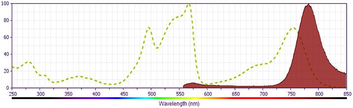
PE-Cy™7 tandem fluorochrome emission is collected in a detector for fluorescence wavelengths of 750 nm and higher. PE-Cy™7-labeled antibodies can be used with FITC- and R-PE-labeled reagents in single-laser flow cytometers with no significant spectral overlap between PE-Cy™7 and FITC.
The OX-35 clone recognizes the CD4 antigen on most thymocytes, a subpopulation of mature T lymphocytes (i.e., MHC class II-restricted T cells, including most T helper cells), monocytes, macrophages, some dendritic cells, and microglia. CD4 is an antigen coreceptor on the T-cell surface that interacts with MHC class II molecules on antigen-presenting cells. It participates in T-cell activation through it's association with the T-cell receptor complex and protein tyrosine kinase Lck. The OX-35 clone has been reported to bind to a different epitope of CD4 than that recognized by the W3/25 and OX-38 clones.
Please refer to Support Documents for Quality Certificates
Global - Refer to manufacturer's instructions for use and related User Manuals and Technical data sheets before using this products as described
Comparisons, where applicable, are made against older BD Technology, manual methods or are general performance claims. Comparisons are not made against non-BD technologies, unless otherwise noted.
For Research Use Only. Not for use in diagnostic or therapeutic procedures.