







Preparation And Storage


Recommended Assay Procedures
For optimal and reproducible results, BD Horizon Brilliant Stain Buffer should be used anytime two or more BD Horizon Brilliant dyes are used in the same experiment. Fluorescent dye interactions may cause staining artifacts which may affect data interpretation. The BD Horizon Brilliant Stain Buffer was designed to minimize these interactions. More information can be found in the Technical Data Sheet of the BD Horizon Brilliant Stain Buffer (Cat. No. 563794).
Product Notices
- Since applications vary, each investigator should titrate the reagent to obtain optimal results.
- An isotype control should be used at the same concentration as the antibody of interest.
- Pacific Blue™ is a trademark of Molecular Probes, Inc., Eugene, OR.
- Source of all serum proteins is from USDA inspected abattoirs located in the United States.
- Caution: Sodium azide yields highly toxic hydrazoic acid under acidic conditions. Dilute azide compounds in running water before discarding to avoid accumulation of potentially explosive deposits in plumbing.
- For fluorochrome spectra and suitable instrument settings, please refer to our Multicolor Flow Cytometry web page at www.bdbiosciences.com/colors.
- Please refer to www.bdbiosciences.com/us/s/resources for technical protocols.
Companion Products





The 13B 1E5 monoclonal antibody specifically binds to C-X-C chemokine receptor type 6 (CXCR6), which is also known as CD186, BONZO, T-lymphocyte-expressed seven-transmembrane domain receptor (TYMSTR), and Seven transmembrane receptor-like from clone 33 (STRL33). CXCR6 is a G-protein coupled chemokine receptor that is expressed on subsets of activated and memory T cells and NKT cells. CXCR6 binds to soluble CXCL16 and membrane-anchored CXCL16 expressed by dendritic cells and macrophages. The CXCL16 and CXCR6 interaction activates Akt and mTor signaling. This regulates cellular migration including the recruitment of tumor-infiltrating lymphocytes and may contribute to the progression and metastasis of various cancers. CXCR6 also can serve as a coreceptor for certain strains of HIV-1 and HIV-2.
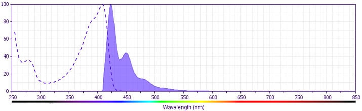
The antibody was conjugated to BD Horizon BV421 which is part of the BD Horizon Brilliant™ Violet family of dyes. With an Ex Max of 407-nm and Em Max at 421-nm, BD Horizon BV421 can be excited by the violet laser and detected in the standard Pacific Blue™ filter set (eg, 450/50-nm filter). BD Horizon BV421 conjugates are very bright, often exhibiting a 10 fold improvement in brightness compared to Pacific Blue conjugates.
Development References (6)
-
Deng HK, Unutmaz D, KewalRamani VN, Littman DR. Expression cloning of new receptors used by simian and human immunodeficiency viruses.. Nature. 1997; 388(6639):296-300. (Biology). View Reference
-
La Porta CA. CXCR6: the role of environment in tumor progression. Challenges for therapy.. Stem Cell Rev. 2012; 8(4):1282-5. (Biology). View Reference
-
Liao F, Alkhatib G, Peden KW, Sharma G, Berger EA, Farber JM. STRL33, A novel chemokine receptor-like protein, functions as a fusion cofactor for both macrophage-tropic and T cell line-tropic HIV-1.. J Exp Med. 1997; 185(11):2015-23. (Biology). View Reference
-
Loetscher M, Amara A, Oberlin E, et al. TYMSTR, a putative chemokine receptor selectively expressed in activated T cells, exhibits HIV-1 coreceptor function.. Curr Biol. 1997; 7(9):652-60. (Biology). View Reference
-
Murphy PM. International Union of Pharmacology. XXX. Update on chemokine receptor nomenclature.. Pharmacol Rev. 2002; 54(2):227-9. (Biology). View Reference
-
Xiao G, Wang X, Wang J, et al. CXCL16/CXCR6 chemokine signaling mediates breast cancer progression by pERK1/2-dependent mechanisms.. Oncotarget. 2015; 6(16):14165-78. (Biology). View Reference
Please refer to Support Documents for Quality Certificates
Global - Refer to manufacturer's instructions for use and related User Manuals and Technical data sheets before using this products as described
Comparisons, where applicable, are made against older BD Technology, manual methods or are general performance claims. Comparisons are not made against non-BD technologies, unless otherwise noted.
For Research Use Only. Not for use in diagnostic or therapeutic procedures.
