
Preparation And Storage
Product Notices
- Since applications vary, each investigator should titrate the reagent to obtain optimal results.
- An isotype control should be used at the same concentration as the antibody of interest.
- Caution: Sodium azide yields highly toxic hydrazoic acid under acidic conditions. Dilute azide compounds in running water before discarding to avoid accumulation of potentially explosive deposits in plumbing.
- Source of all serum proteins is from USDA inspected abattoirs located in the United States.
- Although hamster immunoglobulin isotypes have not been well defined, BD Biosciences Pharmingen has grouped Armenian and Syrian hamster IgG monoclonal antibodies according to their reactivity with a panel of mouse anti-hamster IgG mAbs. A table of the hamster IgG groups, Reactivity of Mouse Anti-Hamster Ig mAbs, may be viewed at http://www.bdbiosciences.com/documents/hamster_chart_11x17.pdf.
- Pacific Blue™ is a trademark of Molecular Probes, Inc., Eugene, OR.
- For fluorochrome spectra and suitable instrument settings, please refer to our Multicolor Flow Cytometry web page at www.bdbiosciences.com/colors.
- Please refer to www.bdbiosciences.com/us/s/resources for technical protocols.
Companion Products


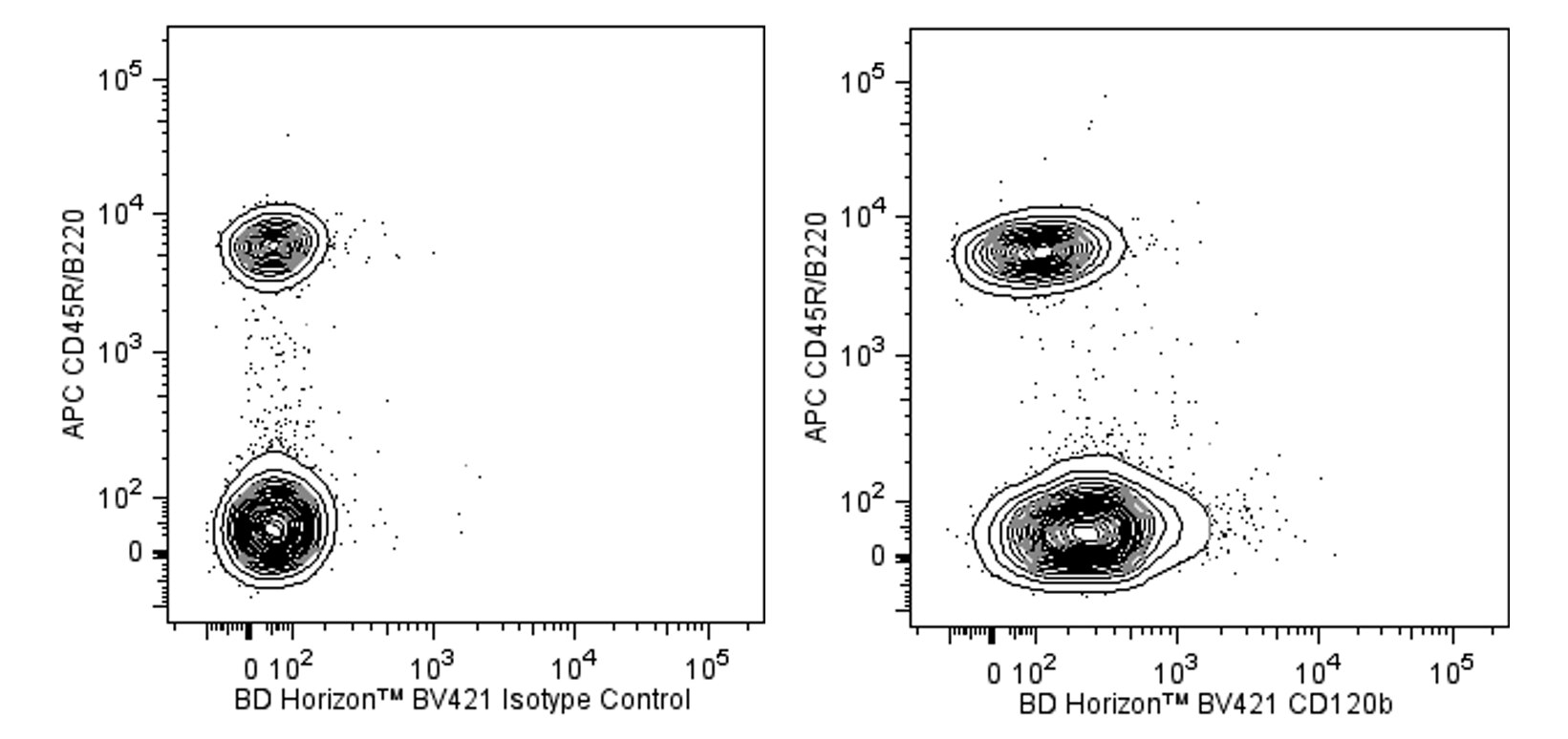
The TR75-89 monoclonal antibody specifically binds to the extracellular region of CD120b, the 75 kDa receptor for the mouse cytokines, tumor necrosis factor (TNF, aka TNF-α) and lymphotoxin-alpha (LT-α, aka lymphotoxin, TNF-β). This receptor, referred to as the p75 or Type II Tumor Necrosis Factor Receptor (TNFRII), or TNFRSF1B, is expressed by a variety of cell lines and normal cell types including T cells, monocytes, macrophages, and neutrophils. Resting B cells express low or undetectable levels of TNFRII whereas mature erythrocytes are uniformly negative for TNFRII expression. In addition, the TR75-89 antibody can bind to a soluble, truncated form of the mouse Type II TNFR that is shed by cells in response to certain stimuli, e.g., cells treated with LPS or TNF. The in vivo administration of nonblocking, nonagonistic TR75-89 antibody reportedly results in the linear accumulation of shed, soluble forms of p75 TNFR in the circulation. The TR75-89 antibody does not recognize the 55 kDa (p55) Type I TNFR (aka, CD120a). TR75-89 does not block the binding and does not neutralize the bioactivity of the TNF ligand on L929 target cell populations that express p75 (and p55) TNFR. The immunogen used to generate the TR75-89 hybridoma was a purified, soluble extracellular domain of the mouse Type II TNFR.
The antibody was conjugated to BD Horizon™ BV421 which is part of the BD Horizon Brilliant™ Violet family of dyes. With an Ex Max of 407-nm and Em Max at 421-nm, BD Horizon BV421 can be excited by the violet laser and detected in the standard Pacific Blue™ filter set (eg, 450/50-nm filter). BD Horizon BV421 conjugates are very bright, often exhibiting a 10 fold improvement in brightness compared to Pacific Blue conjugates.

Development References (3)
-
Pinckard JK, Sheehan KC, Arthur CD, Schreiber RD. Constitutive shedding of both p55 and p75 murine TNF receptors in vivo. J Immunol. 1997; 158(8):3869-3873. (Clone-specific: Bioassay, Immunoprecipitation, Inhibition, In vivo exacerbation). View Reference
-
Pinckard JK, Sheehan KC, Schreiber RD. Ligand-induced formation of p55 and p75 tumor necrosis factor receptor heterocomplexes on intact cells. J Biol Chem. 1997; 272(16):10784-10789. (Clone-specific: Immunoprecipitation). View Reference
-
Sheehan KC, Pinckard JK, Arthur CD, Dehner LP, Goeddel DV, Schreiber RD. Monoclonal antibodies specific for murine p55 and p75 tumor necrosis factor receptors: identification of a novel in vivo role for p75. J Exp Med. 1995; 181(2):607-617. (Immunogen: Immunoprecipitation). View Reference
Please refer to Support Documents for Quality Certificates
Global - Refer to manufacturer's instructions for use and related User Manuals and Technical data sheets before using this products as described
Comparisons, where applicable, are made against older BD Technology, manual methods or are general performance claims. Comparisons are not made against non-BD technologies, unless otherwise noted.
For Research Use Only. Not for use in diagnostic or therapeutic procedures.