
-
Reagents
- Flow Cytometry Reagents
-
Western Blotting and Molecular Reagents
- Immunoassay Reagents
-
Single-Cell Multiomics Reagents
- BD® OMICS-Guard Sample Preservation Buffer
- BD® AbSeq Assay
- BD® OMICS-One Immune Profiler Protein Panel
- BD® Single-Cell Multiplexing Kit
- BD Rhapsody™ ATAC-Seq Assays
- BD Rhapsody™ Whole Transcriptome Analysis (WTA) Amplification Kit
- BD Rhapsody™ TCR/BCR Next Multiomic Assays
- BD Rhapsody™ Targeted mRNA Kits
- BD Rhapsody™ Accessory Kits
-
Functional Assays
-
Microscopy and Imaging Reagents
-
Cell Preparation and Separation Reagents
-
- BD® OMICS-Guard Sample Preservation Buffer
- BD® AbSeq Assay
- BD® OMICS-One Immune Profiler Protein Panel
- BD® Single-Cell Multiplexing Kit
- BD Rhapsody™ ATAC-Seq Assays
- BD Rhapsody™ Whole Transcriptome Analysis (WTA) Amplification Kit
- BD Rhapsody™ TCR/BCR Next Multiomic Assays
- BD Rhapsody™ Targeted mRNA Kits
- BD Rhapsody™ Accessory Kits
- Finland (English)
-
Change country/language
Old Browser
This page has been recently translated and is available in French now.
Looks like you're visiting us from {countryName}.
Would you like to stay on the current country site or be switched to your country?


.png)

Two-color flow cytometric analysis of IL-17A expression in stimulated human peripheral blood lymphocytes. Human peripheral blood mononuclear cells were stimulated for 5 hours with Phorbol 12-Myristate 13-Acetate (Sigma P-8139; 50 ng/ml final concentration) and Ionomycin (Sigma I-0634; 1 μg/ml final concentration) in the presence of BD GolgiStop™ Protein Transport Inhibitor (containing Monensin) (Cat. No. 554724). The cells were harvested, washed with BD Pharmingen™ Stain Buffer (FBS) (Cat. No. 554656), and fixed and permeabilized with BD Cytofix/Cytoperm™ Fixation and Permeabilization Solution (Cat. No. 554722). The cells were then washed and stained in BD Perm/Wash™ Buffer (Cat. No. 554723) with PE Mouse Anti-Human CD4 antibody (Cat. No. 555347/561843/561844) and either BD Horizon™ APC-R700 Mouse IgG1, κ Isotype Control (Cat. No. 564974; Left Plot) or BD Horizon APC-R700 Mouse Anti-Human IL-17A antibody (Cat. No. 565163; Right Plot). Two-color flow cytometric dot plots show the correlated expression patterns of IL-17A (or Ig Isotype control staining) versus CD4 for gated events with the forward and side light-scatter characteristics of intact lymphocytes. Flow cytometric analysis was performed using a BD™ LSR II Flow Cytometer System.
.png)

BD Horizon™ APC-R700 Mouse Anti-Human IL-17A
.png)
Regulatory Status Legend
Any use of products other than the permitted use without the express written authorization of Becton, Dickinson and Company is strictly prohibited.
Preparation And Storage
Product Notices
- This reagent has been pre-diluted for use at the recommended Volume per Test. We typically use 1 × 10^6 cells in a 100-µl experimental sample (a test).
- Please refer to www.bdbiosciences.com/us/s/resources for technical protocols.
- Alexa Fluor® is a registered trademark of Molecular Probes, Inc., Eugene, OR.
- Caution: Sodium azide yields highly toxic hydrazoic acid under acidic conditions. Dilute azide compounds in running water before discarding to avoid accumulation of potentially explosive deposits in plumbing.
- For fluorochrome spectra and suitable instrument settings, please refer to our Multicolor Flow Cytometry web page at www.bdbiosciences.com/colors.
- Source of all serum proteins is from USDA inspected abattoirs located in the United States.
- An isotype control should be used at the same concentration as the antibody of interest.
Companion Products


.png?imwidth=320)
Human IL-17A, also known as IL-17, is a proinflammatory cytokine that is encoded by the IL17A gene in chromosome 6. IL-17A is produced as a disulfide-linked homodimer comprised of two mature 136-amino acid polypeptides. It is a member of the IL-17 family of structurally related cytokines, designated IL-17A through IL-17F. Activated memory T cells, especially Th17 cells (specialized IL-17A-producing CD4+ T cells distinct from Th1 and Th2 cells) produce IL-17 and provide protective immunity against pathogens. Activated CD8+ T cells, γδT cells, NK cells and neutrophils can also be activated to produce IL-17A. IL-17A binds to and exerts its biological activity through IL-17 receptors (IL-17R) that are expressed by a variety of target cells including fibroblasts, epithelial and endothelial cells, monocytes/macrophages and mast cells. The ubiquitous IL-17R expression pattern may explain the broad tissue responsiveness to IL-17. IL-17 induces stromal cells to secrete cytokines and chemokines involved in inflammatory and hematopoietic processes. For example, IL-17 induces fibroblasts to produce IL-6, IL-8, G-CSF and express increased surface ICAM-1. The N49-653 antibody reacts with human IL-17A.
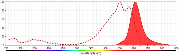
This antibody was conjugated to BD Horizon APC-R700, which has been developed exclusively by BD Biosciences as a better alternative to Alexa Fluor® 700. APC-R700 excites and emits at similar wavelengths to Alexa Fluor® 700 yet exhibits significantly improved brightness. This dye can be excited by the red laser and detected with the same filter set as Alexa Fluor® (eg, 730/45-nm filter).

Development References (9)
-
Boisvert M, Chetoui N, Gendron S, Aoudjit F. Alpha2beta1 integrin is the major collagen-binding integrin expressed on human Th17 cells. Eur J Immunol. 2010; 40(10):2710-2719. (Clone-specific: Flow cytometry). View Reference
-
Fossiez F, Djossou O, Chomarat P, et al. T cell interleukin-17 induces stromal cells to produce proinflammatory and hematopoietic cytokines. J Exp Med. 1996; 183(6):2593-2603. (Biology). View Reference
-
Hong M, Sandalova E, Low D, et al. Trained immunity in newborn infants of HBV-infected mothers.. Nat Commun. 2015; 6:6588. (Clone-specific: Flow cytometry). View Reference
-
Korn T, Oukka M, Kuchroo V, Bettelli E. Th17 cells: effector T cells with inflammatory properties. Semin Immunol. 2007; 19(6):362-371. (Biology). View Reference
-
Melton AC, Melrose J, Alajoki L, et al. Regulation of IL-17A production is distinct from IL-17F in a primary human cell co-culture model of T cell-mediated B cell activation. PLoS ONE. 2013; 8(3):e58966. (Clone-specific: Flow cytometry). View Reference
-
Moseley TA, Haudenschild DR, Rose L, Reddi AH. Interleukin-17 family and IL-17 receptors. Cytokine Growth Factor Rev. 2003; 14(2):155-174. (Biology). View Reference
-
Weaver CT, Hatton RD, Mangan PR, Harrington LE. IL-17 family cytokines and the expanding diversity of effector T cell lineages. Annu Rev Immunol. 2007; 25:821-852. (Biology). View Reference
-
Yao Z, Painter SL, Fanslow WC, et al. Human IL-17: a novel cytokine derived from T cells. J Immunol. 1995; 155(12):5483-5486. (Biology). View Reference
-
Yao Z, Spriggs MK, Derry JM, et al. Molecular characterization of the human interleukin (IL)-17 receptor. Cytokine. 1997; 9(11):794-800. (Biology). View Reference
Please refer to Support Documents for Quality Certificates
Global - Refer to manufacturer's instructions for use and related User Manuals and Technical data sheets before using this products as described
Comparisons, where applicable, are made against older BD Technology, manual methods or are general performance claims. Comparisons are not made against non-BD technologies, unless otherwise noted.
For Research Use Only. Not for use in diagnostic or therapeutic procedures.