

Preparation And Storage
Product Notices
- Since applications vary, each investigator should titrate the reagent to obtain optimal results.
- An isotype control should be used at the same concentration as the antibody of interest.
- Caution: Sodium azide yields highly toxic hydrazoic acid under acidic conditions. Dilute azide compounds in running water before discarding to avoid accumulation of potentially explosive deposits in plumbing.
- The Alexa Fluor®, Pacific Blue™, and Cascade Blue® dye antibody conjugates in this product are sold under license from Molecular Probes, Inc. for research use only, excluding use in combination with microarrays, or as analyte specific reagents. The Alexa Fluor® dyes (except for Alexa Fluor® 430), Pacific Blue™ dye, and Cascade Blue® dye are covered by pending and issued patents.
- Alexa Fluor® is a registered trademark of Molecular Probes, Inc., Eugene, OR.
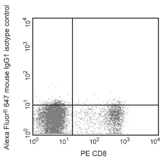
- Alexa Fluor® 647 fluorochrome emission is collected at the same instrument settings as for allophycocyanin (APC).
- For fluorochrome spectra and suitable instrument settings, please refer to our Multicolor Flow Cytometry web page at www.bdbiosciences.com/colors.
- Please refer to www.bdbiosciences.com/us/s/resources for technical protocols.
Companion Products


The SL-12.1 monoclonal antibody specifically recognizes human Interferon regulatory factor 3 (IRF-3). Viral infection in mammals can lead to the induction of multiple pathways as part of the host defense mechanism. One of the major pathways activated is the JAK-STAT pathway by various interferons (IFNα and IFNβ). These IFNs exert their influence via transcriptional activation of specific target genes involved in antiviral defense, for example the chemokine ISG15 gene or the major histocompatibility complex class I and II molecules. These genes in turn are regulated by the JAK-STAT signaling pathway and through interferon regulatory factors (IRFs). IRFs are a family of transcription factors that possess a broad range of activities. IRF-3 is one of nine members which all share a common DNA binding domain which binds to an IFN stimulated response element (ISRE) found in the majority of IFN-inducible promoters. The IRF3 gene expresses a 50 kDa protein which is constitutively expressed in all tissues. The protein undergoes post-translational modification as well as dimerization and is translocated from the cytoplasm to the nucleus upon viral infection or exposure to dsRNA.
Development References (10)
-
Au WC, Moore PA, Lowther W, Juang YT, Pitha PM. Identification of a member of the interferon regulatory factor family that binds to the interferon-stimulated response element and activates expression of interferon-induced genes. Proc Natl Acad Sci U S A. 1995; 92(25):11657-11661. (Biology). View Reference
-
Darnell JE Jr, Kerr IM, Stark GR. Jak-STAT pathways and transcriptional activation in response to IFNs and other extracellular signaling proteins. Science. 1994; 264(5164):1415-1421. (Biology). View Reference
-
Goriely S, Molle C, Nguyen M, et al. Interferon regulatory factor 3 is involved in Toll-like receptor 4 (TLR4)- and TLR3-induced IL-12p35 gene activation.. Blood. 2006; 107(3):1078-84. (Clone-specific: Immunofluorescence). View Reference
-
Hiscott J, Pitha P, Genin P, et al. Triggering the interferon response: the role of IRF-3 transcription factor.. J Interferon Cytokine Res. 1999; 19(1):1-13. (Biology). View Reference
-
Karpova AY, Trost M, Murray JM, Cantley LC, Howley PM. Interferon regulatory factor-3 is an in vivo target of DNA-PK.. Proc Natl Acad Sci USA. 2002; 99(5):2818-23. (Clone-specific: Immunofluorescence, Immunoprecipitation, Western blot). View Reference
-
Loh JE, Chang CH, Fodor WL, Flavell RA. Dissection of the interferon gamma-MHC class II signal transduction pathway reveals that type I and type II interferon systems share common signalling component(s). EMBO J. 1992; 11(4):1351-1363. (Biology). View Reference
-
Mamane Y, Heylbroeck C, Genin P, et al. Interferon regulatory factors: the next generation. Gene. 1999; 237(1):1-14. (Biology). View Reference
-
Reich N, Evans B, Levy D, Fahey D, Knight E Jr, Darnell JE Jr. Interferon-induced transcription of a gene encoding a 15-kDa protein depends on an upstream enhancer element. Proc Natl Acad Sci U S A. 1987; 84(18):6394-6398. (Biology). View Reference
-
Ronco LV, Karpova AY, Vidal M, Howley PM. Human papillomavirus 16 E6 oncoprotein binds to interferon regulatory factor-3 and inhibits its transcriptional activity. Genes Dev. 1998; 12(13):2061-2072. (Immunogen: Immunoprecipitation, Western blot). View Reference
-
Wathelet MG, Lin CH, Parekh BS, Ronco LV, Howley PM, Maniatis T. Virus infection induces the assembly of coordinately activated transcription factors on the IFN-beta enhancer in vivo. Mol Cell. 1998; 1(4):507-518. (Clone-specific: Gel shift, Immunofluorescence, Immunoprecipitation, Western blot). View Reference
Please refer to Support Documents for Quality Certificates
Global - Refer to manufacturer's instructions for use and related User Manuals and Technical data sheets before using this products as described
Comparisons, where applicable, are made against older BD Technology, manual methods or are general performance claims. Comparisons are not made against non-BD technologies, unless otherwise noted.
For Research Use Only. Not for use in diagnostic or therapeutic procedures.