-
Reagents
- Flow Cytometry Reagents
-
Western Blotting and Molecular Reagents
- Immunoassay Reagents
-
Single-Cell Multiomics Reagents
- BD® OMICS-Guard Sample Preservation Buffer
- BD® AbSeq Assay
- BD® Single-Cell Multiplexing Kit
- BD Rhapsody™ ATAC-Seq Assays
- BD Rhapsody™ Whole Transcriptome Analysis (WTA) Amplification Kit
- BD Rhapsody™ TCR/BCR Next Multiomic Assays
- BD Rhapsody™ Targeted mRNA Kits
- BD Rhapsody™ Accessory Kits
- BD® OMICS-One Protein Panels
-
Functional Assays
-
Microscopy and Imaging Reagents
-
Cell Preparation and Separation Reagents
-
- BD® OMICS-Guard Sample Preservation Buffer
- BD® AbSeq Assay
- BD® Single-Cell Multiplexing Kit
- BD Rhapsody™ ATAC-Seq Assays
- BD Rhapsody™ Whole Transcriptome Analysis (WTA) Amplification Kit
- BD Rhapsody™ TCR/BCR Next Multiomic Assays
- BD Rhapsody™ Targeted mRNA Kits
- BD Rhapsody™ Accessory Kits
- BD® OMICS-One Protein Panels
- Finland (English)
-
Change country/language
Old Browser
This page has been recently translated and is available in French now.
Looks like you're visiting us from United States.
Would you like to stay on the current country site or be switched to your country?
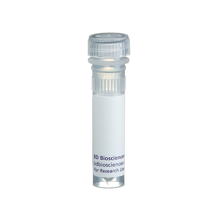
BD Pharmingen™ PI/RNase Staining Buffer

Flow cytometric analysis of DNA content of Jurkat cells. Cells from Jurkat cell line (Human T-cell leukemia; ATCC TIB-152) were fixed with 1% paraformaldehyde (methanol free) and stored in 70% ethanol at -20°C. Cells were stained with 0.5 ml of PI/RNase Staining Buffer (Cat. No. 550825) for 15 minutes at room temperature and analyzed by flow cytometry.


Flow cytometric analysis of DNA content of Jurkat cells. Cells from Jurkat cell line (Human T-cell leukemia; ATCC TIB-152) were fixed with 1% paraformaldehyde (methanol free) and stored in 70% ethanol at -20°C. Cells were stained with 0.5 ml of PI/RNase Staining Buffer (Cat. No. 550825) for 15 minutes at room temperature and analyzed by flow cytometry.

Flow cytometric analysis of DNA content of Jurkat cells. Cells from Jurkat cell line (Human T-cell leukemia; ATCC TIB-152) were fixed with 1% paraformaldehyde (methanol free) and stored in 70% ethanol at -20°C. Cells were stained with 0.5 ml of PI/RNase Staining Buffer (Cat. No. 550825) for 15 minutes at room temperature and analyzed by flow cytometry.


ImageTitle~BD Pharmingen™ PI/RNase Staining Buffer

Regulatory Status Legend
Any use of products other than the permitted use without the express written authorization of Becton, Dickinson and Company is strictly prohibited.
Product Details
Description
Propidium Iodide (PI) is a fluorescent vital dye that stains DNA and RNA. PI binds to both DNA and RNA, so the latter must be removed by digestion with ribonucleases. The content of DNA as determined by flow cytometry, and can reveal useful information about the cell cycle and the proteins involved in cell cycle regulation. Cells in G2 and M phases of the cell cycle have double the DNA content of those in G0 and G1 phases. Cells in S phase have DNA content lying between these extremes. PI is detected in the orange range of the spectrum using a 562-588 nm band pass filter. This reagent may be used to analyze cell cycle by flow cytometry in addition to use with antibodies for examining the expression of proteins during the cell cycle.
Preparation And Storage
Recommended Assay Procedures
Flow cytometry: After fixing and permeabilizing your cell sample, use 0.5 mL /test (1 x 10e6 cells) and incubate for 15 minutes at room temperature before analysis. Please refer to http://static.bdbiosciences.com/documents/BD_FlowCytometry_DNA_Staining_Protocol.pdf for more protocol information.
Product Notices
- Caution: Sodium azide yields highly toxic hydrazoic acid under acidic conditions. Dilute azide compounds in running water before discarding to avoid accumulation of potentially explosive deposits in plumbing.
- Avoid contact with skin and eyes.
- Please refer to http://regdocs.bd.com to access safety data sheets (SDS).
- Please refer to www.bdbiosciences.com/us/s/resources for technical protocols.
Development References (2)
-
Douglas RS, Tarshis AD, Pletcher CH Jr, Nowell PC, Moore JS. A simplified method for the coordinate examination of apoptosis and surface phenotype of murine lymphocytes. J Immunol Methods. 1995; 188(2):219-228. (Biology). View Reference
-
Kalejta RF, Shenk T, Beavis AJ. Use of a membrane-localized green fluorescent protein allows simultaneous identification of transfected cells and cell cycle analysis by flow cytometry. Cytometry. 1997; 29(4):286-291. (Biology). View Reference
Please refer to Support Documents for Quality Certificates
Global - Refer to manufacturer's instructions for use and related User Manuals and Technical data sheets before using this products as described
Comparisons, where applicable, are made against older BD Technology, manual methods or are general performance claims. Comparisons are not made against non-BD technologies, unless otherwise noted.
For Research Use Only. Not for use in diagnostic or therapeutic procedures.