

Preparation And Storage
Recommended Assay Procedures
In human PBMC, overnight serum starvation was found to be necessary for detection of a BMP-6-induced increase in Smad1/8 phosphorylation. Serum starvation for 1 hour following PBMC isolation was not sufficient to reduce basal levels of Smad1 (pS463/pS465)/Smad8(pS465/pS467). Some donor variation was observed in the reduction of basal phosphorylation by overnight serum starvation.
Product Notices
- This reagent has been pre-diluted for use at the recommended Volume per Test. We typically use 1 × 10^6 cells in a 100-µl experimental sample (a test).
- Please refer to www.bdbiosciences.com/us/s/resources for technical protocols.
- Caution: Sodium azide yields highly toxic hydrazoic acid under acidic conditions. Dilute azide compounds in running water before discarding to avoid accumulation of potentially explosive deposits in plumbing.
- Source of all serum proteins is from USDA inspected abattoirs located in the United States.
- For fluorochrome spectra and suitable instrument settings, please refer to our Multicolor Flow Cytometry web page at www.bdbiosciences.com/colors.
Companion Products


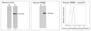
The N6-1233 monoclonal antibody specifically binds to the Smad1 protein phosphorylated at the Ser463/465 sites and the Smad8 protein phosphorylated at the Ser465/467 sites. Smad1 and Smad8 are ~60 kDa proteins and are members of the Smad superfamily. The Smad family members are divided into three subfamilies: receptor regulated Smads or R-Smads, including Smads1, 2, 3, 5, and 8; common partner Smad, or Co-Smad, including Smad4; and inhibitory Smads, or I-Smad, including Smads 6 and 7. Activation of Transforming Growth Factor-beta (TGF-beta) superfamily serine/threonine kinase receptors, such as TGF-beta and Bone Morphogenic Protein (BMP) receptors, leads to the phosphorylation of R-Smads at several sites. It has been shown that Ser463 and Ser465 of Smad1 are phosphorylated by BMP receptors. In B cells and pre-B cells, BMP-6 has been shown to induce Smad1/5/8 phosphorylation and inhibit cell proliferation. Phosphorylated R-Smads form complexes with Co-Smad and translocate into the nucleus to regulate transcription affecting a wide range of critical processes including cell-fate determination, proliferation, morphogenesis, differentiation and apoptosis. The inhibitory Smads inhibit this pathway through two potential mechanisms: either by preventing R-Smads from binding to their corresponding receptors and/or by competing with Smad4, the Co-Smad, from binding to R-Smads. This antibody may crossreact with Smad5 pS463/pS465 based on sequence homology.

Development References (8)
-
Hogan BL. Bone morphogenetic proteins: multifunctional regulators of vertebrate development. Genes Dev. 1996; 10(13):1580-1594. (Biology). View Reference
-
Hoodless PA, Haerry T, Abdollah S, et al. MADR1, a MAD-related protein that functions in BMP2 signaling pathways. Cell. 1996; 85(4):489-500. (Biology). View Reference
-
Kersten C, Dosen G, Myklebust JH, et al. BMP-6 inhibits human bone marrow B lymphopoiesis--upregulation of Id1 and Id3. Exp Hematol. 2006; 34(1):72-81. (Biology). View Reference
-
Kersten C, Sivertsen EA, Hystad ME, Forfang L, Smeland EB, Myklebust JH. BMP-6 inhibits growth of mature human B cells; induction of Smad phosphorylation and upregulation of Id1. BMC Immunol. 2005; 6:9. (Biology). View Reference
-
Kretzschmar M, Doody J, Massagué J. Opposing BMP and EGF signalling pathways converge on the TGF-beta family mediator Smad1. Nature. 1997; 389(6651):618-622. (Biology). View Reference
-
Kretzschmar M, Liu F, Hata A, Doody J, Massague J. The TGF-beta family mediator Smad1 is phosphorylated directly and activated functionally by the BMP receptor kinase. Genes Dev. 1997; 11:984-995. (Biology). View Reference
-
Whitman M. Smads and early developmental signaling by the TGFbeta superfamily. Genes Dev. 1998; 12(16):2445-2462. (Biology). View Reference
-
Yang J, Davies RJ, Southwood M, et al. Mutations in bone morphogenetic protein type II receptor cause dysregulation of Id gene expression in pulmonary artery smooth muscle cells: implications for familial pulmonary arterial hypertension. Circ Res. 2008; 102(10):1212-1221. (Biology). View Reference
Please refer to Support Documents for Quality Certificates
Global - Refer to manufacturer's instructions for use and related User Manuals and Technical data sheets before using this products as described
Comparisons, where applicable, are made against older BD Technology, manual methods or are general performance claims. Comparisons are not made against non-BD technologies, unless otherwise noted.
For Research Use Only. Not for use in diagnostic or therapeutic procedures.