Old Browser
This page has been recently translated and is available in French now.
Looks like you're visiting us from {countryName}.
Would you like to stay on the current country site or be switched to your country?


.png)

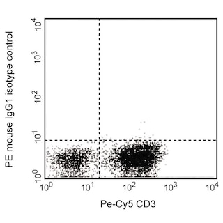
Flow cytometric analysis of N-Cadherin on H9-derived neural stem cells (NSC, left) and transformed human epithelioid carcinoma (HeLa, right). NSC derived from H9 human ES cells (WiCell, Madison, WI) and HeLa cells (ATCC CCL 2.2) were harvested without trypsinization [please note, the epitope is sensitive to trypsin] and stained with either PE Mouse IgG1, κ isotype control (dashed line, Cat. No. 554680) or PE Mouse Mouse Anti-Human CD325 antibody (solid line) at matching concentrations. The histograms were derived from gated events based on light scattering characteristics of the NSC or HeLa cells. Flow cytometry was performed on a BD LSR™ II flow cytometry system.
.png)

BD Pharmingen™ PE Mouse anti-Human CD325
.png)
Regulatory Status Legend
Any use of products other than the permitted use without the express written authorization of Becton, Dickinson and Company is strictly prohibited.
Preparation And Storage
Recommended Assay Procedures
Because the extracellular domain of N-Cadherin is trypsin-sensitive, it is important to avoid using trypsin to dissociate the cells to be studied.
Product Notices
- This reagent has been pre-diluted for use at the recommended Volume per Test. We typically use 1 × 10^6 cells in a 100-µl experimental sample (a test).
- Please refer to www.bdbiosciences.com/us/s/resources for technical protocols.
- An isotype control should be used at the same concentration as the antibody of interest.
- For fluorochrome spectra and suitable instrument settings, please refer to our Multicolor Flow Cytometry web page at www.bdbiosciences.com/colors.
- Source of all serum proteins is from USDA inspected abattoirs located in the United States.
- Caution: Sodium azide yields highly toxic hydrazoic acid under acidic conditions. Dilute azide compounds in running water before discarding to avoid accumulation of potentially explosive deposits in plumbing.
Companion Products


The 8C11 monoclonal antibody recognizes the extracellular domain of human N-Cadherin (CD325). Cadherins are a family of Ca2+ -dependent intercellular adhesion molecules that play a central role in controlling morphogenetic movements during development. Their function is regulated by association with the actin cytoskeleton by a complex of cytoplasmic proteins called the catenins (α, β, γ). Members of the cadherin family include P-cadherin , E-cadherin (uvomorulin), N-cadherin (neural cadherin), R-cadherin, cadherin 5, L-CAM, and EP-cadherin. N-cadherin mRNA is found at elevated levels in brain and heart and at a much lower level in liver. Mechanisms such as mRNA expression, cytokine modulation, and protease-mediated turnover modulate N-cadherin protein levels during development. In addition, N-cadherin function is indirectly regulated by endogenous kinases and phosphatases. Tyrosine phosphorylation of β-catenin complexed with N-cadherin results in dissociation of N-cadherin from actin. However, N-cadherin also interacts with a PTP1B-like phosphatase that dephosphorylates β-catenin and promotes N-cadherin/actin association. Thus, N-cadherin is an integral adhesion molecule whose function is regulated by protein-protein interactions and phosphorylation/dephosphorylation events.

Development References (3)
-
Knudsen KA, Soler AP, Johnson KR, Wheelock MJ. Interaction of alpha-actinin with the cadherin/catenin cell-cell adhesion complex via alpha-catenin. J Cell Biol. 1995; 130:66-77. (Biology). View Reference
-
Puch S, Armeanu S, Kibler C, et al. N-cadherin is developmentally regulated and functionally involved in early hematopoietic cell differentiation. J Cell Sci. 2001; 114(8):1567-1577. (Clone-specific: Flow cytometry). View Reference
-
Wein F, Pietsch L, Saffrich R, et al. N-Cadherin is expressed on human hematopoietic progenitor cells and mediates interaction with human mesenchymal stromal cells. Stem Cell Res. 2010; 4(2):129-139. (Clone-specific: Flow cytometry). View Reference
Please refer to Support Documents for Quality Certificates
Global - Refer to manufacturer's instructions for use and related User Manuals and Technical data sheets before using this products as described
Comparisons, where applicable, are made against older BD Technology, manual methods or are general performance claims. Comparisons are not made against non-BD technologies, unless otherwise noted.
For Research Use Only. Not for use in diagnostic or therapeutic procedures.
Report a Site Issue
This form is intended to help us improve our website experience. For other support, please visit our Contact Us page.