







Preparation And Storage


Recommended Assay Procedures
This antibody conjugate is suitable for intracellular staining of human peripheral blood mononuclear cells (using BD Cytofix™ Fixation Buffer). Any of the three BD Phosflow™ permeabilization buffers may be used.
Product Notices
- This reagent has been pre-diluted for use at the recommended Volume per Test. We typically use 1 × 10^6 cells in a 100-µl experimental sample (a test).
- Please refer to www.bdbiosciences.com/us/s/resources for technical protocols.
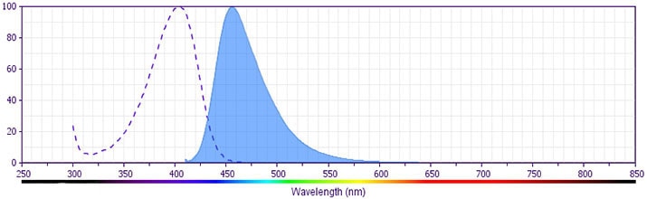
- Pacific Blue™ has a maximum absorption of 416 nm and maximum emission of 451 nm. Before staining with this reagent, please confirm that your flow cytometer is capable of exciting the fluorochrome and discriminating the resulting fluorescence.
- For fluorochrome spectra and suitable instrument settings, please refer to our Multicolor Flow Cytometry web page at www.bdbiosciences.com/colors.
- The Alexa Fluor®, Pacific Blue™, and Cascade Blue® dye antibody conjugates in this product are sold under license from Molecular Probes, Inc. for research use only, excluding use in combination with microarrays, or as analyte specific reagents. The Alexa Fluor® dyes (except for Alexa Fluor® 430), Pacific Blue™ dye, and Cascade Blue® dye are covered by pending and issued patents.
- Caution: Sodium azide yields highly toxic hydrazoic acid under acidic conditions. Dilute azide compounds in running water before discarding to avoid accumulation of potentially explosive deposits in plumbing.
- Source of all serum proteins is from USDA inspected abattoirs located in the United States.
- Pacific Blue™ is a trademark of Molecular Probes, Inc., Eugene, OR.
- All other brands are trademarks of their respective owners.
Companion Products

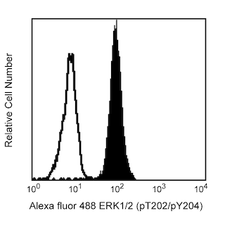

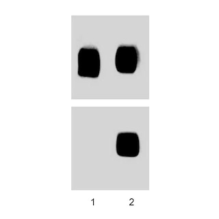
Activation of the immune and inflammatory responses often involves the recognition of bacterial endotoxin (lipopolysaccharide or LPS). Binding of LPS by monocytes results in the production and release of proinflammatory cytokines, such as IL-1 and TNF. LPS-induced signaling cascades involve members of the Ser/Thr protein kinase family known as the Mitogen Activated Protein Kinases (MAPKs). MAPK signal transduction pathways mediate the effects of various extracellular stimuli on biological processes such as proliferation, differentiation, and death. The p38 MAPKs include p38α (MAPK14), β (MAPK11), γ (MAPK12), and δ (MAPK13). These Ser/Thr kinases are activated by dual phosphorylation on threonine (T) and tyrosine (Y) within the motif Thr-Gly-Tyr located in kinase subdomain VIII. Activation of p38 MAPK is mediated specifically by the MAP Kinase Kinases, MKK3, MKK4, and MKK6. This leads to the activation of multiple transcription factors (NF-κB, ATF-2, Elk-1, and CHOP) that induce expression of many different genes, including proinflammatory cytokine genes. Thus, p38 MAPKs are central kinases in multiple signal transduction pathways.
The 36/p38 (pT180/pY182) monoclonal antibody recognizes the conserved dual phosphorylated site pT180/pY182 of p38α, β, γ, and δ.
Development References (3)
-
Brunet A, Pouyssegur J. Identification of MAP kinase domains by redirecting stress signals into growth factor responses. Science. 1996; 272(5268):1652-1655. (Biology). View Reference
-
Han J, Lee JD, Bibbs L, Ulevitch RJ. A MAP kinase targeted by endotoxin and hyperosmolarity in mammalian cells. Science. 1994; 265(5173):808-811. (Biology). View Reference
-
Winston BW, Chan ED, Johnson GL, Riches DW. Activation of p38mapk, MKK3, and MKK4 by TNF-alpha in mouse bone marrow-derived macrophages. J Immunol. 1997; 159(9):4491-4497. (Biology). View Reference
Please refer to Support Documents for Quality Certificates
Global - Refer to manufacturer's instructions for use and related User Manuals and Technical data sheets before using this products as described
Comparisons, where applicable, are made against older BD Technology, manual methods or are general performance claims. Comparisons are not made against non-BD technologies, unless otherwise noted.
For Research Use Only. Not for use in diagnostic or therapeutic procedures.
