Old Browser
This page has been recently translated and is available in French now.
Looks like you're visiting us from {countryName}.
Would you like to stay on the current country site or be switched to your country?


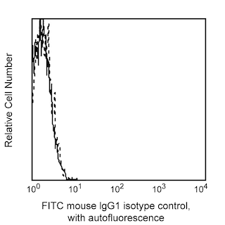
Flow cytometric analysis of CD94 expression on human peripheral blood lymphocytes. Human whole blood was stained with either FITC Mouse Anti-Human CD94 (Cat. No. 555888; solid line histogram) or FITC Mouse IgG1, κ Isotype Control (Cat. No. 555748; dashed line histogram). Erythrocytes were lysed with BD Pharm Lyse™ Lysing Buffer (Cat. No. 555899). Fluorescent histograms were derived from gated events with the side and forward light-scattering characteristics of viable lymphocytes. Flow cytometry was performed on a BD FACScan™ system.


BD Pharmingen™ FITC Mouse Anti-Human CD94

Regulatory Status Legend
Any use of products other than the permitted use without the express written authorization of Becton, Dickinson and Company is strictly prohibited.
Preparation And Storage
Product Notices
- This reagent has been pre-diluted for use at the recommended Volume per Test. We typically use 1 × 10^6 cells in a 100-µl experimental sample (a test).
- An isotype control should be used at the same concentration as the antibody of interest.
- Caution: Sodium azide yields highly toxic hydrazoic acid under acidic conditions. Dilute azide compounds in running water before discarding to avoid accumulation of potentially explosive deposits in plumbing.
- Source of all serum proteins is from USDA inspected abattoirs located in the United States.
- For fluorochrome spectra and suitable instrument settings, please refer to our Multicolor Flow Cytometry web page at www.bdbiosciences.com/colors.
- Please refer to www.bdbiosciences.com/us/s/resources for technical protocols.
Companion Products


The HP-3D9 monoclonal antibody specifically binds to CD94 which is also known as KP43. CD94 is expressed on the cell surface as a ~70 kDa, disulfide-linked, type II transmembrane glycoprotein dimer. It is encoded by KLRD1 (Killer cell lectin like receptor D1) which belongs to the C-type lectin superfamily. CD94 is expressed on natural killer (NK) cells, especially activated NK cells. It is also expressed on γ/δ TCR+ T lymphocytes, NK-T cells, and on some CD8+CD56+ α/β TCR+ cells. CD94 associates with various NKG2 receptors to form receptors for HLA class I molecules and plays a role in regulating cellular adhesion and activation. The HP-3D9 antibody can reportedly inhibit the cytolytic activity of activated NK cells.

Development References (3)
-
Aramburu J, Balboa MA, Ramírez A, et al. A novel functional cell surface dimer (Kp43) expressed by natural killer cells and T cell receptor-gamma/delta+ T lymphocytes. I. Inhibition of the IL-2-dependent proliferation by anti-Kp43 monoclonal antibody. J Immunol. 1990; 144(8):3238-3247. (Biology). View Reference
-
Balboa MA, Balsinde J, Aramburu J, Mollinedo F, López-Botet M. Phospholipase D activation in human natural killer cells through the Kp43 and CD16 surface antigens takes place by different mechanisms. Involvement of the phospholipase D pathway in tumor necrosis factor alpha synthesis. J Exp Med. 1992; 176(1):9-17. (Biology). View Reference
-
Pérez-Villar JJ, Melero I, Rodríguez A, et al. Functional ambivalence of the Kp43 (CD94) NK cell-associated surface antigen. J Immunol. 1995; 154(11):5779-5788. (Biology). View Reference
Please refer to Support Documents for Quality Certificates
Global - Refer to manufacturer's instructions for use and related User Manuals and Technical data sheets before using this products as described
Comparisons, where applicable, are made against older BD Technology, manual methods or are general performance claims. Comparisons are not made against non-BD technologies, unless otherwise noted.
For Research Use Only. Not for use in diagnostic or therapeutic procedures.
Report a Site Issue
This form is intended to help us improve our website experience. For other support, please visit our Contact Us page.