The site is undergoing maintenance. Ordering can continue through fax and phone.
Sunday, August 16, 2026, 1:30 - 4:30 pm (CEST).
If you encounter problems while ordering. Please contact us at BDBCustomerService@bd.com
See More
The site is undergoing maintenance. Ordering can continue through fax and phone.
Sunday, August 16, 2026, 1:30 - 4:30 pm (CEST).
If you encounter problems while ordering. Please contact us at BDBCustomerService@bd.com
See MoreThe site is undergoing maintenance. Ordering can continue through fax and phone.
Sunday, August 16, 2026, 1:30 - 4:30 pm (CEST)
.If you encounter problems while ordering. Please contact us at BDBCustomerService@bd.com
This page has been recently translated and is available in French now.
Looks like you're visiting us from {countryName}.
Would you like to stay on the current location site or be switched to your location?
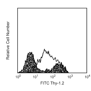
Multiparameter flow cytometric analysis using BD OptiBuild™ BV786 Rat Anti-Mouse CD274 antibody (Cat. No. 741014; right plot)) on live mouse splenocytes stimulated by plate-bound NA/LE Hamster Anti-Mouse CD3e antibody for 3 days, with FMO control (left plot). Flow cytometry was performed using a BD LSRFortessa™ X-20 Flow Cytometer System.
Any use of products other than the permitted use without the express written authorization of Becton, Dickinson and Company is strictly prohibited.
For optimal and reproducible results, BD Horizon Brilliant Stain Buffer should be used anytime two or more BD Horizon Brilliant dyes (including BD OptiBuild Brilliant reagents) are used in the same experiment. Fluorescent dye interactions may cause staining artifacts which may affect data interpretation. The BD Horizon Brilliant Stain Buffer was designed to minimize these interactions. More information can be found in the Technical Data Sheet of the BD Horizon Brilliant Stain Buffer (Cat. No. 563794).
The MIH5 monoclonal antibody specifically binds to CD274, also known as B7-H1 or PDL1, a 43-kDa glycoprotein encoded by the Pdcd1lg1 gene of the B7 family of the Ig superfamily. Pdcd1lg1 mRNA is expressed in more tissues than other members of the B7 family; transcripts are found in lymphoid tissues and many, but not all, non-lymphoid tissues. The protein has been detected at low levels on resting peripheral T and B lymphocytes, macrophages, and dendritic cells. B7-H1 mRNA and protein expression are upregulated upon activation of T and B cells, macrophages, dendritic cells, and epidermal keratinocytes by a variety of stimulatory factors. B7-H1's receptor, PD-1, contains an ITIM (Immunoreceptor Tyrosine-based Inhibitory Motif) on its intracytoplasmic region and is expressed on activated B and T lymphocytes, suggesting that B7-H1-PD-1 interaction may be involved in the negative regulation of immune responses. The second PD-1 ligand, B7-DC (PD-L2), is also a member of the B7 family of the Ig superfamily. Furthermore, B7-H1 may participate in positive immunoregulation, or costimulation of T cells, through an additional receptor, which is not PD-1 and distinct from the alternate receptor for B7-DC. The MIH5 antibody blocks the binding of PD-1-Ig to B7-H1 transfectants.
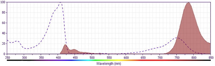
The antibody was conjugated to BD Horizon™ BV786 which is part of the BD Horizon Brilliant™ Violet family of dyes. This dye is a tandem fluorochrome of BD Horizon BV421 with an Ex Max of 405-nm and an acceptor dye with an Em Max at 786-nm. BD Horizon BV786 can be excited by the violet laser and detected in a filter used to detect Cy™7-like dyes (eg, 780/60-nm filter).
Please refer to Support Documents for Quality Certificates
Global - Refer to manufacturer's instructions for use and related User Manuals and Technical data sheets before using this products as described
Comparisons, where applicable, are made against older BD Technology, manual methods or are general performance claims. Comparisons are not made against non-BD technologies, unless otherwise noted.
For Research Use Only. Not for use in diagnostic or therapeutic procedures.