The site is undergoing maintenance. Ordering can continue through fax and phone.
Sunday, August 16, 2026, 1:30 - 4:30 pm (CEST).
If you encounter problems while ordering. Please contact us at BDBCustomerService@bd.com
See More
The site is undergoing maintenance. Ordering can continue through fax and phone.
Sunday, August 16, 2026, 1:30 - 4:30 pm (CEST).
If you encounter problems while ordering. Please contact us at BDBCustomerService@bd.com
See MoreThe site is undergoing maintenance. Ordering can continue through fax and phone.
Sunday, August 16, 2026, 1:30 - 4:30 pm (CEST)
.If you encounter problems while ordering. Please contact us at BDBCustomerService@bd.com
This page has been recently translated and is available in French now.
Looks like you're visiting us from {countryName}.
Would you like to stay on the current location site or be switched to your location?
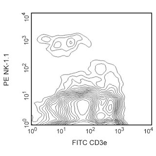
Multicolor flow cytometric analysis of CD49b expression on mouse splenocytes. Splenic leucocytes from a C57BL6/J mouse were stained with PE Mouse Anti-Mouse NK1.1 antibody (Cat. No. 553165/557391/561046) and either BD Horizon™ BV421 Rat IgM, κ Isotype Control (Cat. No. 562708, Left Panel) or BD Horizon™ BV421 Rat Anti-Mouse CD49b antibody (Cat. No. 563063, Right Panel). Two color flow cytometric dot plots showing the expression CD49b (or Ig Isotype control staining) versus NK1.1 were derived from gated events with the forward and side light-scatter characteristics of viable splenocytes. Flow cytometry was performed using a BD™ LSR II Flow Cytometer System.
Any use of products other than the permitted use without the express written authorization of Becton, Dickinson and Company is strictly prohibited.
The rat anti-mouse CD49b monoclonal antibody (clone DX5) specifically binds to the integrin α2 chain (CD49b). CD49b is a 150 kDa transmembrane glycoprotein that non-covalently associates with CD29 (integrin β1) to form the integrin α2β1 complex known as VLA-2. The rat anti-mouse CD49b antibody (clone DX5) has been reported to identify the majority of NK cells and a small T-cell subpopulation in most mouse strains (e.g., A/J, AKR, BALB/c, C3H/HeJ, C57BL/6, C57BL/10, C57BR, C58, CBA/Ca, DBA/1, DBA/2, SJL, SWR, 129/J, but not NOD). The DX5 antibody also recognizes platelets that express high levels of CD49b. Multiparameter flow cytometric analysis has demonstrated that most lymphocytes which express NK-1.1 (NKR-P1B and NKR-P1C), as detectable by mouse anti-mouse NK-1.1 antibody (clone PK136), also express the DX5 antigen. Small DX5+ NK-1.1- and DX5- NK-1.1+ cell subsets are found, especially among the CD3-positive cell population. Some CD49b+ NK cells have been reported to gradually lose reactivity with the rat anti-mouse CD49b antibody (clone DX5) when cultured in the presence of recombinant human IL-2. The resulting DX5-negative cells have weakened cytotoxic activity when compared to the remaining DX5+ cells. This indicates that the DX5 antibody distinguishes functional subsets of NK cells. No activation or blocking activity of the rat anti-mouse antibody (clone DX5) has been observed. Staining of splenic NK cells with this antibody reportedly can be blocked by hamster anti-mouse CD49b antibody (clone HMα2).
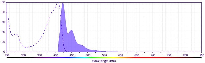
The antibody was conjugated to BD Horizon™ BV421 which is part of the BD Horizon™ Brilliant Violet™ family of dyes. With an Ex Max of 407-nm and Em Max at 421-nm, BD Horizon™ BV421 can be excited by the violet laser and detected in the standard Pacific Blue™ filter set (eg, 450/50-nm filter). BD Horizon™ BV421 conjugates are very bright, often exhibiting a 10 fold improvement in brightness compared to Pacific Blue™ conjugates.
Please refer to Support Documents for Quality Certificates
Global - Refer to manufacturer's instructions for use and related User Manuals and Technical data sheets before using this products as described
Comparisons, where applicable, are made against older BD Technology, manual methods or are general performance claims. Comparisons are not made against non-BD technologies, unless otherwise noted.
For Research Use Only. Not for use in diagnostic or therapeutic procedures.