-
Reagents
- Flow Cytometry Reagents
-
Western Blotting and Molecular Reagents
- Immunoassay Reagents
-
Single-Cell Multiomics Reagents
- BD® OMICS-Guard Sample Preservation Buffer
- BD® AbSeq Assay
- BD® Single-Cell Multiplexing Kit
- BD Rhapsody™ ATAC-Seq Assays
- BD Rhapsody™ Whole Transcriptome Analysis (WTA) Amplification Kit
- BD Rhapsody™ TCR/BCR Next Multiomic Assays
- BD Rhapsody™ Targeted mRNA Kits
- BD Rhapsody™ Accessory Kits
- BD® OMICS-One Protein Panels
-
Functional Assays
-
Microscopy and Imaging Reagents
-
Cell Preparation and Separation Reagents
Old Browser
This page has been recently translated and is available in French now.
Looks like you're visiting us from {countryName}.
Would you like to stay on the current location site or be switched to your location?
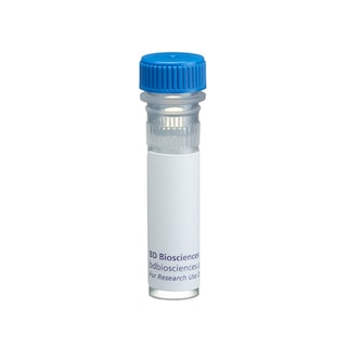
BD Transduction Laboratories™ Purified Mouse Anti-Human MSH3
Clone 52/MSH3 (RUO)


Western blot analysis of MSH3 on a HeLa cell lysate (Human cervical epitheloid carcinoma; ATCC CCL-2). 2 µg/mL (lane 1), 1 µg/mL (lane 2) and 0.5 µg/mL (lane 3) of the mouse anti-human MSH3 antibody were used.

Immunofluorescence staining of human fibroblasts.


Regulatory Status Legend
Any use of products other than the permitted use without the express written authorization of Becton, Dickinson and Company is strictly prohibited.
Preparation And Storage
Recommended Assay Procedures
Western blot: Please refer to http://www.bdbiosciences.com/pharmingen/protocols/Western_Blotting.shtml
Product Notices
- Since applications vary, each investigator should titrate the reagent to obtain optimal results.
- Source of all serum proteins is from USDA inspected abattoirs located in the United States.
- Caution: Sodium azide yields highly toxic hydrazoic acid under acidic conditions. Dilute azide compounds in running water before discarding to avoid accumulation of potentially explosive deposits in plumbing.
- Please refer to www.bdbiosciences.com/us/s/resources for technical protocols.
Companion Products


Bacterial mismatch DNA repair involves the MutL, MutH, and MutS proteins, which forms a complex that mediates excision repair. Mutations in or deficiencies of any of these proteins results in a mutator phenotype that is characterized by genetic instability. Human homologs of MutS include MSH2, MSH3, and MSH6. MSH2 forms heterodimers with MSH6 (hMutSα) or MSH3 (hMutSβ) that specifically bind single-mispaired nucleotides and a subset of insertion-deletion mismatches. In addition, these heterodimers have intrinsic ATPase activity that is regulated by mismatch binding. ADP-bound heterodimers bind mismatched nucleotides, while ATP-bound heterodimers do not. The role of MSH3 in genetic stability in human cells in unclear. However, MSH3 and MSH6 share roles in the control of mutation rates. Both participate in repair of replication errors containing base-base mismatches or 1-4 extra bases. The MSH3 gene is located upstream of the dihydrofolate reductase (DHFR) gene and is expressed at low levels in a variety of human tissues. Thus, MSH3 is a component of an adenosine nucleotide-regulated molecular switch whose activity is essential for classical nucleotide mismatch repair.
Development References (4)
-
New L, Liu K, Crouse GF. The yeast gene MSH3 defines a new class of eukaryotic MutS homologues. Mol Gen Genet. 1993; 239(1-2):97-108. (Biology). View Reference
-
Umar A, Risinger JI, Glaab WE, Tindall KR, Barrett JC, Kunkel TA. Functional overlap in mismatch repair by human MSH3 and MSH6. Genetics. 1998; 148(4):1637-1646. (Biology). View Reference
-
Watanabe A, Ikejima M, Suzuki N, Shimada T. Genomic organization and expression of the human MSH3 gene. Genomics. 1996; 31(3):311-318. (Biology). View Reference
-
Wilson T, Guerrette S, Fishel R. Dissociation of mismatch recognition and ATPase activity by hMSH2-hMSH3. J Biol Chem. 1999; 274(31):21659-21664. (Biology). View Reference
Please refer to Support Documents for Quality Certificates
Global - Refer to manufacturer's instructions for use and related User Manuals and Technical data sheets before using this products as described
Comparisons, where applicable, are made against older BD Technology, manual methods or are general performance claims. Comparisons are not made against non-BD technologies, unless otherwise noted.
For Research Use Only. Not for use in diagnostic or therapeutic procedures.