





BD Pharmingen™ Alexa Fluor® 488 Mouse anti-Human Nanog
Clone N31-355 (RUO)


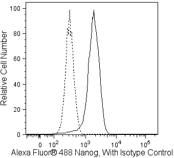
Alexa Fluor® 488 anti-human Nanog staining of human embryonic stem cells. H9 cells (WiCell, Madison, WI), passage 34, were harvested, fixed in BD Cytofix™ buffer (Cat. No. 554655), permeabilized with BD™ Phosflow Perm/Wash buffer I (Cat. No. 557885) and stained with Alexa Fluor® 488 anti-human Nanog (Cat. No. 560791, solid line) or Alexa Fluor® 488 Mouse IgG1, κ Isotype Control (Cat. No.557702, dashed line). Flow cytometry was performed on a BD™ LSR II flow cytometry system. This reagent will also work in BD™ Phosflow Perm II and III buffers (Cat. No. 558052,558050 respectively).
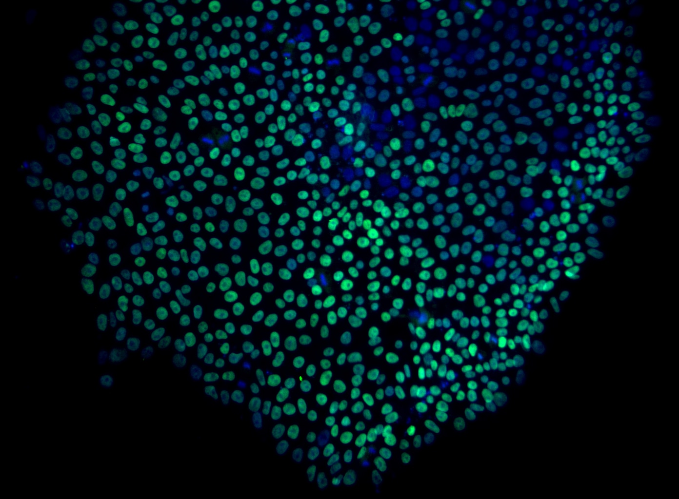
Immunoflourescent staining of human embryonic stem cells with Alexa Fluor® 488 anti-human Nanog. H9 cells (WiCell, Madison, WI), passage 45, grown in mTESR™1 media (StemCell Technologies) on BD Matrigel™ hESC-qualified Matrix (Cat. No. 354277) were fixed in BD Cytofix™ buffer, permeabilized, and stained with Alexa Fluor® 488 anti-Human Nanog monoclonal antibody (pseudo-colored green) at 2.5 µg/mL. Cell nuclei were counter-stained with Hoechst 33342 (Cat. No. 561908, pseudo-colored blue). The images were captured on a BD Pathway™ 435 Cell Analyzer and merged using BD Attovision™ Software. Permeabilization was done using BD™ Phosflow Perm/Wash buffer I for this antibody; Triton™ X-100 is also suitable for permeabilization.

Alexa Fluor® 488 anti-human Nanog staining of human embryonic stem cells. H9 cells (WiCell, Madison, WI), passage 34, were harvested, fixed in BD Cytofix™ buffer (Cat. No. 554655), permeabilized with BD™ Phosflow Perm/Wash buffer I (Cat. No. 557885) and stained with Alexa Fluor® 488 anti-human Nanog (Cat. No. 560791, solid line) or Alexa Fluor® 488 Mouse IgG1, κ Isotype Control (Cat. No.557702, dashed line). Flow cytometry was performed on a BD™ LSR II flow cytometry system. This reagent will also work in BD™ Phosflow Perm II and III buffers (Cat. No. 558052,558050 respectively).
Immunoflourescent staining of human embryonic stem cells with Alexa Fluor® 488 anti-human Nanog. H9 cells (WiCell, Madison, WI), passage 45, grown in mTESR™1 media (StemCell Technologies) on BD Matrigel™ hESC-qualified Matrix (Cat. No. 354277) were fixed in BD Cytofix™ buffer, permeabilized, and stained with Alexa Fluor® 488 anti-Human Nanog monoclonal antibody (pseudo-colored green) at 2.5 µg/mL. Cell nuclei were counter-stained with Hoechst 33342 (Cat. No. 561908, pseudo-colored blue). The images were captured on a BD Pathway™ 435 Cell Analyzer and merged using BD Attovision™ Software. Permeabilization was done using BD™ Phosflow Perm/Wash buffer I for this antibody; Triton™ X-100 is also suitable for permeabilization.


Preparation And Storage


Product Notices
- This reagent has been pre-diluted for use at the recommended Volume per Test. We typically use 1 × 10^6 cells in a 100-µl experimental sample (a test).
- Source of all serum proteins is from USDA inspected abattoirs located in the United States.
- Caution: Sodium azide yields highly toxic hydrazoic acid under acidic conditions. Dilute azide compounds in running water before discarding to avoid accumulation of potentially explosive deposits in plumbing.
- The Alexa Fluor®, Pacific Blue™, and Cascade Blue® dye antibody conjugates in this product are sold under license from Molecular Probes, Inc. for research use only, excluding use in combination with microarrays, or as analyte specific reagents. The Alexa Fluor® dyes (except for Alexa Fluor® 430), Pacific Blue™ dye, and Cascade Blue® dye are covered by pending and issued patents.
- Alexa Fluor® 488 fluorochrome emission is collected at the same instrument settings as for fluorescein isothiocyanate (FITC).
- Alexa Fluor® is a registered trademark of Molecular Probes, Inc., Eugene, OR.
- Triton is a trademark of the Dow Chemical Company.
- For fluorochrome spectra and suitable instrument settings, please refer to our Multicolor Flow Cytometry web page at www.bdbiosciences.com/colors.
- Please refer to www.bdbiosciences.com/us/s/resources for technical protocols.
The N31-355 monoclonal antibody reacts with human Nanog (named for Tir Na Nog, the land of the ever-young of Celtic mythology), which is a homeobox transcription factor required for the maintenance of the undifferentiated state of pluripotent stem cells. Nanog expression counteracts the differentiation-promoting signals induced by the extrinsic factors LIF (Leukemia Inhibitory Factor) and BMP (Bone Morphogenic Protein). When Nanog expression is down-regulated, cell differentiation can proceed. Proteins that regulate Nanog expression include transcription factors Oct4, SOX2, FoxD3, and Tcf3 and tumor suppressor p53. Nanog is one of the factors that can contribute to reprogramming of differentiated cells to an induced pluripotent stem cell state.

Development References (7)
-
Chambers I, Colby D, Robertson M, et al. Functional expression cloning of Nanog, a pluripotency sustaining factor in embryonic stem cells. Cell. 2003; 113:643-655. (Biology). View Reference
-
Ezeh UI, Turek PJ, Reijo RA, Clark AT. Human embryonic stem cell genes OCT4, NANOG, STELLAR, and GDF3 are expressed in both seminoma and breast carcinoma. Cancer. 2005; 104(10):2255-2265. (Biology). View Reference
-
Mitsui K, Tokuzawa Y, Itoh H, et al. The homeoprotein Nanog is required for maintenance of pluripotency in mouse epiblast and ES cells. Cell. 2003; 113:631-642. (Biology). View Reference
-
Pan G, Thomson JA. Nanog and transcriptional networks in embryonic stem cell pluripotency. Cell Res. 2007; 17:42-49. (Biology). View Reference
-
Sun Y, Li H, Yang H, Rao MS, Zhan M. Mechanisms controlling embryonic stem cell self-renewal and differentiation. Crit Rev Eukaryot Gene Expr.. 2006; 16(3):211-231. (Biology). View Reference
-
Suzuki A, Raya A, Kawakami Y, et al. Nanog binds to Smad1 and blocks bone morphogenetic protein-induced differentiation of embryonic stem cells. Proc Natl Acad Sci U S A. 2006; 103(27):10294-10299. (Biology). View Reference
-
Yu J, Vodyanik MA, Smuga-Otto K, et al. Induced pluripotent stem cell lines derived from human somatic cells. Science. 2007; 318(5858):1917-1920. (Biology). View Reference
Please refer to Support Documents for Quality Certificates
Global - Refer to manufacturer's instructions for use and related User Manuals and Technical data sheets before using this products as described
Comparisons, where applicable, are made against older BD Technology, manual methods or are general performance claims. Comparisons are not made against non-BD technologies, unless otherwise noted.
For Research Use Only. Not for use in diagnostic or therapeutic procedures.