
Preparation And Storage
Recommended Assay Procedures
Flow cytometry: Chemokine receptors are known to internalize during manipulation resulting in low frequency expression. Investigators are advised to perform immunophenotyping studies of chemokine receptors on freshly collected samples (<24 Hrs). Incubation with the antibody should be done at 4°C in the dark. Cellular manipulation, such as Ficoll separation, freezing, or exposure to cold temperatures prior to staining should be minimized and have been shown to cause a decrease in staining intensity and/or inconsistent results.
Investigators should note that alternative staining procedures may be necessary. A multiple-step staining procedure is strongly recommended, in some instances, to amplify immunofluorescent signals for the flow cytometric analysis of mouse CXCR5 expression. Investigators may find the Purified Rat Anti-Mouse CXCR5 antibody (Cat. No. 551961) to be useful in conjunction with appropriate secondary and tertiary reagents for detecting low frequency expression, such as with Biotin Mouse Anti-Rat IgG2a (Cat. No. 553894) and PE Streptavidin (Cat. No. 554061) or FITC Streptavidin (Cat. No. 554060).
Product Notices
- Since applications vary, each investigator should titrate the reagent to obtain optimal results.
- An isotype control should be used at the same concentration as the antibody of interest.
- Caution: Sodium azide yields highly toxic hydrazoic acid under acidic conditions. Dilute azide compounds in running water before discarding to avoid accumulation of potentially explosive deposits in plumbing.
- This APC-conjugated reagent can be used in any flow cytometer equipped with a dye, HeNe, or red diode laser.
- For fluorochrome spectra and suitable instrument settings, please refer to our Multicolor Flow Cytometry web page at www.bdbiosciences.com/colors.
- Please refer to http://regdocs.bd.com to access safety data sheets (SDS).
- Please refer to www.bdbiosciences.com/us/s/resources for technical protocols.
Companion Products
.png?imwidth=320)


.png?imwidth=320)
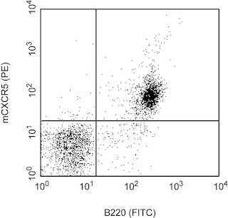
The 2G8 monoclonal antibody specifically binds to the mouse C-X-C Chemokine Receptor type 5, CXCR5. CXCR5 is also known as CD185, BLR1, NLR and MDR15. CXCR5 is a seven-transmembrane, G-protein-coupled receptor that is specific for the CXC chemokine, CXCL13/BLC/BCA-1. The expression of CXCR5 has been detected in spleen, lymph nodes, tonsils, brain, bone marrow, T cells, B cells, cerebrum, cerebellum, hippcampus and pituitary. In mouse spleen, CXCR5 was strictly expressed by mature B cells and a small subset of T lymphocytes. CXCR5 plays a role in directing the migration of B and T cells to B cell follicles with the spleen and certain other lymphoid tissues. The immunogen used to generate 2G8 hybridoma was a recombinant protein containing N-terminal amino acids of mouse CXCR5 (GST-NmBLR1).

Development References (7)
-
Barella L, Loetscher M, Tobler A, Baggiolini M, Moser B. Sequence variation of a novel heptahelical leucocyte receptor through alternative transcript formation. Biochem J. 1995; 309(3):773-779. (Biology). View Reference
-
Dobner T, Wolf I, Emrich T, Lipp M. Differentiation-specific expression of a novel G protein-coupled receptor from Burkitt's lymphoma. Eur J Immunol. 1992; 22(11):2795-2799. (Biology). View Reference
-
Forster R, Mattis AE, Kremmer E, Wolf E, Brem G, Lipp M. A putative chemokine receptor, BLR1, directs B cell migration to defined lymphoid organs and specific anatomic compartments of the spleen. Cell. 1996; 87(6):1037-1047. (Immunogen). View Reference
-
Gunn MD, Ngo VN, Ansel KM, Ekland EH, Cyster JG, Williams LT. A B-cell-homing chemokine made in lymphoid follicles activates Burkitt's lymphoma receptor-1. Nature. 1998; 391(6669):799-803. (Biology). View Reference
-
Kaiser E, Forster R, Wolf I, Ebensperger C, Kuehl WM, Lipp M. The G protein-coupled receptor BLR1 is involved in murine B cell differentiation and is also expressed in neuronal tissues. Eur J Immunol. 1993; 23(10):2532-2539. (Biology). View Reference
-
Kouba M, Vanetti M, Wang X, Schafer M, Hollt V. Cloning of a novel putative G-protein-coupled receptor (NLR) which is expressed in neuronal and lymphatic tissue. FEBS Lett. 1993; 321(2-3):173-178. (Biology). View Reference
-
Legler DF, Loetscher M, Roos RS, Clark-Lewis I, Baggiolini M, Moser B. B cell-attracting chemokine 1, a human CXC chemokine expressed in lymphoid tissues, selectively attracts B lymphocytes via BLR1/CXCR5. J Exp Med. 1998; 187(4):655-660. (Biology). View Reference
Please refer to Support Documents for Quality Certificates
Global - Refer to manufacturer's instructions for use and related User Manuals and Technical data sheets before using this products as described
Comparisons, where applicable, are made against older BD Technology, manual methods or are general performance claims. Comparisons are not made against non-BD technologies, unless otherwise noted.
For Research Use Only. Not for use in diagnostic or therapeutic procedures.