-
Reagents
- Flow Cytometry Reagents
-
Western Blotting and Molecular Reagents
- Immunoassay Reagents
-
Single-Cell Multiomics Reagents
- BD® OMICS-Guard Sample Preservation Buffer
- BD® AbSeq Assay
- BD® Single-Cell Multiplexing Kit
- BD Rhapsody™ ATAC-Seq Assays
- BD Rhapsody™ Whole Transcriptome Analysis (WTA) Amplification Kit
- BD Rhapsody™ TCR/BCR Next Multiomic Assays
- BD Rhapsody™ Targeted mRNA Kits
- BD Rhapsody™ Accessory Kits
- BD® OMICS-One Protein Panels
-
Functional Assays
-
Microscopy and Imaging Reagents
-
Cell Preparation and Separation Reagents
-
- BD® OMICS-Guard Sample Preservation Buffer
- BD® AbSeq Assay
- BD® Single-Cell Multiplexing Kit
- BD Rhapsody™ ATAC-Seq Assays
- BD Rhapsody™ Whole Transcriptome Analysis (WTA) Amplification Kit
- BD Rhapsody™ TCR/BCR Next Multiomic Assays
- BD Rhapsody™ Targeted mRNA Kits
- BD Rhapsody™ Accessory Kits
- BD® OMICS-One Protein Panels
- Belgium (English)
-
Change country/language
Old Browser
This page has been recently translated and is available in French now.
Looks like you're visiting us from United States.
Would you like to stay on the current country site or be switched to your country?
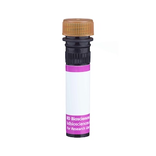
BD Pharmingen™ AKP Streptavidin


Regulatory Status Legend
Any use of products other than the permitted use without the express written authorization of Becton, Dickinson and Company is strictly prohibited.
Preparation And Storage
Recommended Assay Procedures
Immunohistochemical Preparation and Staining Procedure for Frozen Sections
Materials needed:
Phosphate-buffered saline (PBS); 2-methylbutane (isopentane); Acetone; Liquid nitrogen; Dry ice; Hydrogen peroxide (H2O2); Graded alcohols; Xylene
Peel-A-Way® Embedding Molds (Polysciences Inc., Warrington, PA).
OCT Compound for tissue freezing and embedding material (Tissue-Tek®; Sakura Finetek USA).
BCIP®/NBT (5-Bromo-4-Chloro-3-Indolyl Phosphate/ p-Iodonitrotetrazolium) Liquid Substrate System (Sigma-Aldrich).
I. Fixation, Processing, and Sectioning of Tissue for Frozen Sections
1. Label embedding mold and partially fill the mold with OCT Compound.
2. Place fresh tissue sample in pre-labeled embedding molds.
3. Plunge embedding mold with tissue into 2-methylbutane prechilled in a dewar of liquid nitrogen until the block ALMOST solidifies (30 seconds). NOTE: If the block is left in too long, it may crack.
4. Remove tissue block from 2-methylbutane using long forceps.
5. Place blocked tissues on dry ice. (Tissues may be stored in the embedding molds.)
6. Store frozen tissue blocks in -70°C freezer until sectioning.
7. For sectioning, attach the frozen tissue block on the cryostat chuck by adhering it with a small amount of frozen tissue matrix and allow to freeze.
8. Routine sections are cut at 5 microns and picked up on a glass slide, eg, Superfrost® Plus Microscope Slides.
10. Dry overnight at room temperature (RT).
11. Fix sections in cold acetone (-20°C) for 2 min or other suitable fixative (eg, alcohol, formal alcohol, formalin, etc.).
12. Dry fixed slides completely [usually 1 hour at room temperature (RT)].
13. Store in a -70°C freezer until use.
II. Standard Immunohistochemical Staining Procedure for Frozen Sections
Perform all incubations in a humid chamber and do not allow sections to dry out. Isotype and system controls should also be run and must be matched to the isotype of each primary antibody to be tested.
Materials needed:
Phosphate-buffered saline (PBS)
BCIP®/NBT Liquid Substrate System (Sigma-Aldrich)
Please read entire procedure before staining slides.
1. Remove frozen slides prepared in advance from the freezer and allow to come to RT.
2. Label all slides with a pen that uses solvent resistant ink and demarcate the tissue if required.
3. Rinse slides 2-3 times in PBS to remove frozen mounting media.
4. Apply a 0.03% H2O2 in PBS solution (~10 min) to block endogenous peroxidase activity.
5. Rinse slides with one change PBS.
6. Wipe excess buffer from around the specimen.
7. Block with 5% normal serum diluted in PBS for 15 min.*
8. Apply primary antibody diluted in BD Pharmingen™ Antibody Diluent for IHC (Cat. No. 559148) to cover tissue sections on slide and incubate 1 hr at RT in a humid chamber.
9. Rinse slides in 3 changes of PBS, 2 min each.
Optional: Block endogenous biotin using an Endogenous Biotin Blocking Buffer, eg, Biotin/Avidin Blocking Kit (Vector Laboratories, Cat. No. SP-2001) if tissue has endogenous biotin that may cause background staining.
10. Wipe slide again and apply biotinylated secondary antibody diluted in BD Pharmingen™ Antibody Diluent for IHC and allow to incubate at RT for 30 min. This antibody must be matched to recognize the species and isotype of the primary antibody.
11. Rinse slides in 3 changes of PBS, 2 min each.
12. Wipe again and apply AKP Strepavidin using BD Pharmingen™ AKP Streptavidin (Cat. No. 551008) to each slide and incubate at RT 30 min.
13. Rinse slides in 3 changes of PBS, 2 min each.
14. Prepare BCIP/NBT as a chromogenic substrate for the alkaline phosphatase. BCIP/NBT is a widely used chromogen for immunohistochemical staining, eg, BCIP®/NBT Liquid Substrate System (Sigma-Aldrich).
15. Drain PBS from slides, place them on a flat surface, and apply the BCIP/NBT substrate solution making sure all the section is covered by the solution. Allow slides to incubate and check the slide for color development after 5 min or until the desired color intensity is obtained. AKP acts on BCIP/NBT to generate an insoluble NBT diformazan end product that is blue to purple in color. This end product is soluble in alcohol and therefore must be used with an aqueous counterstain and mounting media.
16. Rinse slides well in water 3 times.
17. Counterstain tissues as desired.
18. Add 1-2 drops of a commercially available mounting medium (eg, Aqua-Mount® Mounting Medium) on the tissue and place a glass coverslip over the tissue followed by sealing (eg, with clear nail polish) if desired.
NOTES:
* Always use serum from the species in which the secondary antibody is made: ie, if secondary antibody is Goat Anti-Mouse Ig, then block with 5% normal goat serum. Incubate for 10-30 min before application of the primary antibody. Do not rinse after this step (tap off the blocking solution if using humid chambers) and go straight to the primary antibody application.
Immunohistochemical Preparation and Staining Procedure for Paraffin-Embedded Sections
Materials needed:
Phosphate-buffered saline (PBS); Commercially available: 10% Neutral Buffered Formalin for IHC; Hydrogen peroxide (H2O2); Graded alcohols; Xylene; Hematoxylin.
I. Fixation and Processing of Tissue for Paraffin Sections.
Please read entire procedure before staining slides.
A. Fixation of Tissues in 10% Neutral Buffered Formalin
1. Tissues to be fixed and processed should be cut to a size no larger than 3 mm thick. Let tissues fix in commercially available 10% Neutral Buffered Formalin at RT for 8 hours but not to exceed 24 hours.
2. Follow processing schedule recommended in section C.
B. Alternative Fixation of Tissues in Zinc Fixatives:
1. Many antigenic epitopes are masked or even destroyed by 10% formalin fixation. In some cases, fixation in a milder fixative such as BD Pharmingen™ IHC Zinc Fixative (Cat. No. 550523) or BD Pharmingen™ 10X Zinc Fixative (Formalin Free) [Cat. No. 552658] is helpful to preserve the antigenic epitopes. Place fresh tissues trimmed 3 mm thick into fixative and allow tissues to fix for 24-48 hours at RT.
2. Follow processing schedule recommended in section C.
C. Processing Schedule: Note: The processing, embedding, and sectioning of paraffin blocks requires highly specialized equipment and expertise and is usually performed by a histology or pathology laboratory. While hand processing can be performed according to the following protocol, the results may show marked variation is histology quality and antigenicity.
Station Time Solution
1 Delay Treat Tissue with Fixative
2 45 min 70% Alcohol
3 45 min 80% Alcohol
4 45 min 95% Alcohol
5 45 min 100% Alcohol
6 60 min 100% Alcohol
7 60 min 100% Alcohol
8 60 min Clearing Reagent (xylene or substitute)
9 60 min Clearing Reagent (xylene or substitute)
10 60 min Paraffin 1
11 60 min Paraffin 2
12 60 min Paraffin 3
II. Preparation of Slides with Paraffin Sections for Immunohistochemistry
A. Sectioning and Preparation of Slides.
1. Section paraffin blocks at the desired thickness (usually 4-5 μm) on a microtome and float on a water bath containing deionized or distilled water.
2. Sections are picked up on a glass slide, eg, Superfrost® Plus Microscope Slides.
B. Deparaffinization and Re-hydration of Tissue Slides:
1. Before deparaffinization, place the slides in a 55°C oven for 10 min to melt the paraffin. Deparaffinize slides in 2 changes of xylene or xylene substitute for 5 min each.
2. Transfer slides to 100% alcohol, 2 changes for 3 min each and transfer once through 95% alcohol for 3 min.
3. Block endogenous peroxidase activity by incubating sections in 3% H2O2 solution in methanol for 10 min.
4. Rinse in PBS 2 times for 5 min each time.
Optional: If the antigen of interest is altered during the fixation process, then an antigen retrieval method can be applied. For antigen retrieval to unmask the antigenic epitope use the reagents and protocols detailed in BD Pharmingen™ Retrievagen A (pH 6.0) [Cat. No. 550524] or BD Pharmingen™ BD Retrievagen B (pH 9.5) [Cat. No. 550527].
5. Block with 5% normal serum.
6. Dilute primary antibody in Antibody Diluent for IHC (Cat. No. 559148) to cover tissue sections on slide and incubate at 4°C overnight in a humid chamber.
7. Next day, remove the slides from the refrigerator (4°C) and start with Step 9 of the Standard Immunohistochemical Staining Procedure.
Observe the color of the antibody staining in the tissue sections under microscopy.
Product Notices
- Please refer to www.bdbiosciences.com/us/s/resources for technical protocols.
- Source of all serum proteins is from USDA inspected abattoirs located in the United States.
- Since applications vary, each investigator should titrate the reagent to obtain optimal results.
- Caution: Sodium azide yields highly toxic hydrazoic acid under acidic conditions. Dilute azide compounds in running water before discarding to avoid accumulation of potentially explosive deposits in plumbing.
- Please refer to http://regdocs.bd.com to access safety data sheets (SDS).
- For U.S. patents that may apply, see bd.com/patents.
Data Sheets
Companion Products


Recently Viewed
Streptavidin is a non-glycosylated protein that is purified chromatographically from the bacterium Streptomyces avidinii. Streptavidin homotetramers have a particularly high, non-covalent binding affinity for biotin. When conjugated with fluorochromes, streptavidin has been widely used with biotin-conjugated primary or secondary antibodies and other biotinylated specific-binding molecules (eg, recombinant proteins and lectins) to stain cells and tissues for subsequent multiparameter analysis by flow cytometry, fluorescence microscopy and imaging. When conjugated with an enzyme such as Alkaline Phosphatase (AKP) and coupled with the use of a colorimetric, luminescent, or fluorescent substrate development system, AKP Streptavidin has found widespread use along with biotinylated primary or secondary antibodies in a number of applications including Western blot, ELISA, ELISPOT, Immunocytochemistry and Immunohistochemistry.
Development References (6)
-
Beckstead JH. A simple technique for preservation of fixation-sensitive antigens in paraffin-embedded tissues. J Histochem Cytochem. 1994; 42(8):1127-1134. (Methodology: Immunocytochemistry, Immunohistochemistry). View Reference
-
Chan JK. Advances in immunohistochemical techniques: toward making things simpler, cheaper, more sensitive, and more reproducible. Adv Anat Pathol. 1998; 5(5):314-325. (Methodology: Immunocytochemistry, Immunohistochemistry). View Reference
-
Goldstein M, Watkins S. Immunohistochemistry.. Curr Protoc Mol Biol. 2008; Chapter 14:Unit 14.6. (Methodology: Immunocytochemistry, Immunohistochemistry). View Reference
-
Hofman FM, Taylor CR. Immunohistochemistry.. Curr Protoc Immunol. 2013; 103:21.4.1-21.4.26. (Methodology: Immunocytochemistry, Immunohistochemistry). View Reference
-
Nitta, H., W. Munger, E. Wilson, R. Ralston, and H. Alila. Improved in situ immunodetection of leukocytes on paraffin-embedded mouse spleen. Cell Vision. 1997; 4:314-325. (Methodology: Immunocytochemistry, Immunohistochemistry).
-
Shi SR, Key ME, Kalra KL. Antigen retrieval in formalin-fixed, paraffin-embedded tissues: an enhancement method for immunohistochemical staining based on microwave oven heating of tissue sections. J Histochem Cytochem. 1991; 39(6):741-748. (Methodology: Immunocytochemistry, Immunohistochemistry). View Reference
Please refer to Support Documents for Quality Certificates
Global - Refer to manufacturer's instructions for use and related User Manuals and Technical data sheets before using this products as described
Comparisons, where applicable, are made against older BD Technology, manual methods or are general performance claims. Comparisons are not made against non-BD technologies, unless otherwise noted.
For Research Use Only. Not for use in diagnostic or therapeutic procedures.
