Old Browser
This page has been recently translated and is available in French now.
Looks like you're visiting us from {countryName}.
Would you like to stay on the current country site or be switched to your country?


Left Figure: Western blot analysis of cyclin B1. Lane 1: K562 human leukemia cell lysate. Lane 2: 293 human embryonic kidney cell lysate. Anti-human cyclin B1 (Cat. No. 554176) identifies cyclin B1 as an ~62 kDa band. Right Figure: Cyclin B1 staining of U-2 OS (ATCC HTB-96) cells. Cells were seeded in a 96 well imaging plate (Cat. No. 353219) at ~10,000 cells per well. After overnight incubation, cells were stained using the alcohol perm protocol and the anti-cyclin B1 antibody. The second step reagent was Alexa Fluor® 488 goat anti mouse Ig (Invitrogen). Images were taken on a BD Pathway™ 855 Bioimager system using a 20x objective. This antibody also stained A549 (ATCC CCL-185) and HeLa (CCL-2) cells and worked with both the Triton™ X-100 and alcohol perm protocols (see Recommended Assay Procedure).


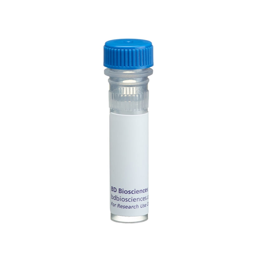
BD Pharmingen™ Purified Mouse Anti-Cyclin B1

Regulatory Status Legend
Any use of products other than the permitted use without the express written authorization of Becton, Dickinson and Company is strictly prohibited.
Preparation And Storage
Recommended Assay Procedures
Bioimaging
1. Seed the cells in appropriate culture medium at ~10,000 cells per well in a BD Falcon™ 96-well Imaging Plate (Cat. No. 353219) and culture overnight.
2. Remove the culture medium from the wells, and fix the cells by adding 100 μl of BD Cytofix™ Fixation Buffer (Cat. No. 554655) to each well. Incubate for 10 minutes at room temperature (RT).
3. Remove the fixative from the wells, and permeabilize the cells using either BD Perm Buffer III, 90% methanol, or Triton™ X-100:
a. Add 100 μl of -20°C 90% methanol or Perm Buffer III (Cat. No. 558050) to each well and incubate for 5 minutes at RT.
OR b. Add 100 μl of 0.1% Triton™ X-100 to each well and incubate for 5 minutes at RT.
4. Remove the permeabilization buffer, and wash the wells twice with 100 μl of 1× PBS.
5. Remove the PBS, and block the cells by adding 100 μl of BD Pharmingen™ Stain Buffer (FBS) (Cat. No. 554656) to each well. Incubate for 30 min at RT.
6. Remove the blocking buffer and add 50 μl of the optimally titrated primary antibody (diluted in Stain Buffer) to each well, and incubate for 1 hr at RT.
7. Remove the primary antibody, and wash the wells three times with 100 μl of 1× PBS.
8. Remove the PBS, and add the second step reagent at its optimally titrated concentration in 50 μl to each well, and incubate in dark for 1 hr at RT.
9. Remove the second step reagent, and wash the wells three times with 100 μl of 1× PBS.
10. Remove the PBS, and counter-stain the nuclei by adding 200 μl per well of 2 μg/ml Hoechst 33342 (e.g., Sigma-Aldrich Cat. No. B2261) in 1× PBS to each well at least 15 min before imaging.
11. View and analyze the cells on an appropriate imaging instrument.
Bioimaging: For more detailed information please refer to http://www.bdbiosciences.com/support/resources/protocols/ceritifed_reagents.jsp
Western blot: For more detailed information please refer to http://www.bdbiosciences.com/pharmingen/protocols/Western_Blotting.shtml
Product Notices
- Since applications vary, each investigator should titrate the reagent to obtain optimal results.
- Please refer to www.bdbiosciences.com/us/s/resources for technical protocols.
- This antibody has been developed and certified for the bioimaging application. However, a routine bioimaging test is not performed on every lot. Researchers are encouraged to titrate the reagent for optimal performance.
- Caution: Sodium azide yields highly toxic hydrazoic acid under acidic conditions. Dilute azide compounds in running water before discarding to avoid accumulation of potentially explosive deposits in plumbing.
- Triton is a trademark of the Dow Chemical Company.
Companion Products


Cyclins and cyclin-dependent kinases (cdks) are evolutionarily conserved proteins that are essential for cell-cycle control in eukaryotes. Cyclins (regulatory subunits) bind to cdks (catlytic subunits) to form complexes that regulate the progression of the cell cycle. The main cyclin-cdks complexes formed in vertebrate cells are cyclin D-cdk4 (G0/G1), cyclin E-cdk2 (G1/S), cyclin A-cdk2 (S) and cyclin B1-cdk1 (G2/M). These complexes are regulated by activating and inhibitory phosphorylation events, as well as by interactions with small regulatory proteins, such as p21 and p27 [Kip1]. Cyclin B1 is a mitotic cyclin, where expression is normally low in G0/G1, increases in S and is maximal during the G2/M phase. Cyclin B1 is rapidly degraded at the end of mitosis, and is required for cells to exit from mitosis. This antibody has been reported to react to hamster and mouse cyclin B1. In addition, the GNS-1 antibody has been reported to recognize an epitope between amino acids 1-21 of human cyclin B1.
Development References (13)
-
Cao L, Faha B, Dembski M, Tsai LH, Harlow E, Dyson N. Independent binding of the retinoblastoma protein and p107 to the transcription factor E2F. Nature. 1992; 355(6356):176-179. (Clone-specific). View Reference
-
Coleman TR, Tang Z, Dunphy WG. Negative regulation of the wee1 protein kinase by direct action of the nim1/cdr1 mitotic inducer. Cell. 1993; 72(6):919-929. (Clone-specific: Western blot). View Reference
-
Darzynkiewicz Z, Gong J, Juan G, Ardelt B, Traganos F. Cytometry of cyclin proteins. Cytometry. 1996; 25(1):1-13. (Methodology: Flow cytometry).
-
Faha B, Ewen ME, Tsai LH, Livingston DM, Harlow E. Interaction between human cyclin A and adenovirus E1A-associated p107 protein. Science. 1992; 255(5040):87-90. (Biology). View Reference
-
Faha B, Harlow E, Lees E. The adenovirus E1A-associated kinase consists of cyclin E-p33cdk2 and cyclin A-p33cdk2. J Virol. 1993; 67(5):2456-2465. (Clone-specific: Immunoprecipitation). View Reference
-
Gong J, Ardelt B, Traganos F, Darzynkiewicz Z. Unscheduled expression of cyclin B1 and cyclin E in several leukemic and solid tumor cell lines. Cancer Res. 1994; 54(16):4285-4288. (Clone-specific: Flow cytometry). View Reference
-
Gong J, Traganos F, Darzynkiewicz Z. Discrimination of G2 and mitotic cells by flow cytometry based on different expression of cyclins A and B1. Exp Cell Res. 1995; 220(1):226-231. (Clone-specific: Flow cytometry, Western blot). View Reference
-
Gong J, Traganos F, Darzynkiewicz Z. Simultaneous analysis of cell cycle kinetics at two different DNA ploidy levels based on DNA content and cyclin B measurements. Cancer Res. 1993; 53(21):5096-5099. (Clone-specific: Flow cytometry).
-
Gong J, Traganos F, and Darzynkiewicz Z. Expression of cyclins B and E in individual MOLT-4 cells and in stimulated human lymphocytes during progression through the cell cycle. Int J Oncol. 1993; 3:1037-1042. (Clone-specific: Flow cytometry).
-
Kung AL, Sherwood SW, Schimke RT. Differences in the regulation of protein synthesis, cyclin B accumulation, and cellular growth in response to the inhibition of DNA synthesis in Chinese hamster ovary and HeLa S3 cells. J Biol Chem. 1993; 268(31):23072-23080. (Clone-specific: Flow cytometry). View Reference
-
Sherwood SW, Kung AL, Roitelman J, Simoni RD, Schimke RT. In vivo inhibition of cyclin B degradation and induction of cell-cycle arrest in mammalian cells by the neutral cysteine protease inhibitor N-acetylleucylleucylnorleucinal. Proc Natl Acad Sci U S A. 1993; 90(8):3353-3357. (Clone-specific: Fluorescence microscopy, Immunohistochemistry, Western blot). View Reference
-
Sherwood SW, Rush DF, Kung AL, Schimke RT. Cyclin B1 expression in HeLa S3 cells studied by flow cytometry. Exp Cell Res. 1994; 211(2):275-281. (Clone-specific: Flow cytometry, Fluorescence microscopy, Immunohistochemistry). View Reference
-
Zhang H, Xiong Y, Beach D. Proliferating cell nuclear antigen and p21 are components of multiple cell cycle kinase complexes. Mol Biol Cell. 1993; 4(9):897-906. (Clone-specific: Flow cytometry, Immunoprecipitation). View Reference
Please refer to Support Documents for Quality Certificates
Global - Refer to manufacturer's instructions for use and related User Manuals and Technical data sheets before using this products as described
Comparisons, where applicable, are made against older BD Technology, manual methods or are general performance claims. Comparisons are not made against non-BD technologies, unless otherwise noted.
For Research Use Only. Not for use in diagnostic or therapeutic procedures.
Report a Site Issue
This form is intended to help us improve our website experience. For other support, please visit our Contact Us page.