
Preparation And Storage
Product Notices
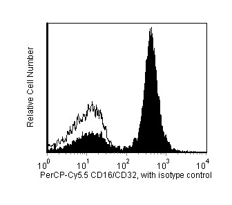
- Since applications vary, each investigator should titrate the reagent to obtain optimal results.
- An isotype control should be used at the same concentration as the antibody of interest.
- Please observe the following precautions: Absorption of visible light can significantly alter the energy transfer occurring in any tandem fluorochrome conjugate; therefore, we recommend that special precautions be taken (such as wrapping vials, tubes, or racks in aluminum foil) to prevent exposure of conjugated reagents, including cells stained with those reagents, to room illumination.
- This PerCP-conjugated product is sold under license to the following patent: US Patent No. 4,876,190.
- Cy is a trademark of Amersham Biosciences Limited. This conjugated product is sold under license to the following patents: US Patent Nos. 5,486,616; 5,569,587; 5,569,766; 5,627,027.
- This product is subject to proprietary rights of Amersham Biosciences Corp. and Carnegie Mellon University and made and sold under license from Amersham Biosciences Corp. This product is licensed for sale only for research. It is not licensed for any other use. If you require a commercial license to use this product and do not have one return this material, unopened to BD Biosciences, 10975 Torreyana Rd, San Diego, CA 92121 and any money paid for the material will be refunded.
- PerCP-Cy5.5 is optimized for use with a single argon ion laser emitting 488-nm light. Because of the broad absorption spectrum of the tandem fluorochrome, extra care must be taken when using dual-laser cytometers, which may directly excite both PerCP and Cy5.5™. We recommend the use of cross-beam compensation during data acquisition or software compensation during data analysis.
- PerCP-Cy5.5–labelled antibodies can be used with FITC- and R-PE–labelled reagents in single-laser flow cytometers with no significant spectral overlap of PerCP-Cy5.5, FITC, and R-PE fluorescence.
- Caution: Sodium azide yields highly toxic hydrazoic acid under acidic conditions. Dilute azide compounds in running water before discarding to avoid accumulation of potentially explosive deposits in plumbing.
- For fluorochrome spectra and suitable instrument settings, please refer to our Multicolor Flow Cytometry web page at www.bdbiosciences.com/colors.
- Please refer to www.bdbiosciences.com/us/s/resources for technical protocols.

The 2.4G2 antibody specifically recognizes a common nonpolymorphic epitope on the extracellular domains of the mouse FcγIII (CD16) and FcγII (CD32) Receptors. It has also been reported to bind the FcγI receptor (CD64) via its Fc domain. 2.4G2 mAb blocks non-antigen-specific binding of immunoglobulins to the FcγIII and FcγII, and possibly FcγI, Receptors in vitro and in vivo. CD16 and/or CD32 are expressed on natural killer cells, monocytes, macrophages, dendritic cells (at low levels), Kupffer cells, granulocytes, mast cells, B lymphocytes, immature thymocytes, and some activated mature T lymphocytes.

Development References (19)
-
Araujo-Jorge T, Rivera MT, el Bouhdidi A, Daeron M, Carlier Y. An Fc gamma RII-, Fc gamma RIII-specific monoclonal antibody (2.4G2) decreases acute Trypanosoma cruzi infection in mice. Infect Immun. 1993; 61(11):4925-4928. (Biology: Blocking). View Reference
-
Benhamou M, Bonnerot C, Fridman WH, Daeron M. Molecular heterogeneity of murine mast cell Fc gamma receptors. J Immunol. 1990; 144(8):3071-3077. (Biology: Immunoprecipitation). View Reference
-
Jensen WA, Marschner S, Ott VL, Cambier JC. FcgammaRIIB-mediated inhibition of T-cell receptor signal transduction involves the phosphorylation of SH2-containing inositol 5-phosphatase (SHIP), dephosphorylation of the linker of activated T-cells (LAT) and inhibition of calcium mobilization. Biochem Soc Trans. 2001; 29(6):840-846. (Biology: Blocking). View Reference
-
Kaji K, Takeshita S, Miyake K, Takai T, Kudo A. Functional association of CD9 with the Fc gamma receptors in macrophages. J Immunol. 2001; 166(5):3256-3265. (Biology: (Co)-stimulation). View Reference
-
Katz HR, Arm JP, Benson AC, Austen KF. Maturation-related changes in the expression of Fc gamma RII and Fc gamma RIII on mouse mast cells derived in vitro and in vivo. J Immunol. 1990; 145(10):3412-3417. (Biology: Immunoprecipitation). View Reference
-
Kurlander RJ, Ellison DM, Hall J. The blockade of Fc receptor-mediated clearance of immune complexes in vivo by a monoclonal antibody (2.4G2) directed against Fc receptors on murine leukocytes. J Immunol. 1984; 133(2):855-862. (Biology: Blocking). View Reference
-
Latour S, Bonnerot C, Fridman WH, Daeron M. Induction of tumor necrosis factor-alpha production by mast cells via Fc gamma R. Role of the Fc gamma RIII gamma subunit. J Immunol. 1992; 149(6):2155-2162. (Biology: (Co)-stimulation). View Reference
-
Lewis VA, Koch T, Plutner H, Mellman I. A complementary DNA clone for a macrophage-lymphocyte Fc receptor. Nature. 1986; 324(6095):372-375. (Biology). View Reference
-
Maeda K, Burton GF, Padgett DA, et al. Murine follicular dendritic cells and low affinity Fc receptors for IgE (Fc epsilon RII). J Immunol. 1992; 148(8):2340-2347. (Biology: Immunohistochemistry). View Reference
-
Mellman IS, Unkeless JC. Purificaton of a functional mouse Fc receptor through the use of a monoclonal antibody. J Exp Med. 1980; 152(4):1048-1069. (Biology: Immunoprecipitation). View Reference
-
Perussia B, Tutt MM, Qiu WQ, et al. Murine natural killer cells express functional Fc gamma receptor II encoded by the Fc gamma R alpha gene. J Exp Med. 1989; 170(1):73-86. (Biology). View Reference
-
Ravetch JV, Luster AD, Weinshank R, et al. Structural heterogeneity and functional domains of murine immunoglobulin G Fc receptors. Science. 1986; 234(4777):718-725. (Biology). View Reference
-
Rodewald HR, Awad K, Moingeon P, et al. Fc gamma RII/III and CD2 expression mark distinct subpopulations of immature CD4-CD8- murine thymocytes: in vivo developmental kinetics and T cell receptor beta chain rearrangement status. J Exp Med. 1993; 177(4):1079-1092. (Biology: Immunoprecipitation). View Reference
-
Rodewald HR, Moingeon P, Lucich JL, Dosiou C, Lopez P, Reinherz EL. A population of early fetal thymocytes expressing Fc gamma RII/III contains precursors of T lymphocytes and natural killer cells. Cell. 1992; 69(1):139-150. (Biology: Immunoprecipitation). View Reference
-
Takezawa R, Watanabe Y, Akaike T. Direct evidence of macrophage differentiation from bone marrow cells in the liver: a possible origin of Kupffer cells. J Biochem (Tokyo). 1995; 118(6):1175-1183. (Biology). View Reference
-
Titus JA, Finkelman FD, Stephany DA, Jones JF, Segal DM. Quantitative analysis of Fc gamma receptors on murine spleen cell populations by using dual parameter flow cytometry. J Immunol. 1984; 133(2):556-561. (Biology). View Reference
-
Unkeless JC. Characterization of a monoclonal antibody directed against mouse macrophage and lymphocyte Fc receptors. J Exp Med. 1979; 150(3):580-596. (Immunogen). View Reference
-
Vremec D, Zorbas M, Scollay R, et al. The surface phenotype of dendritic cells purified from mouse thymus and spleen: investigation of the CD8 expression by a subpopulation of dendritic cells. J Exp Med. 1992; 176(1):47-58. (Biology). View Reference
-
Witmer MD, Steinman RM. The anatomy of peripheral lymphoid organs with emphasis on accessory cells: light-microscopic immunocytochemical studies of mouse spleen, lymph node, and Peyer's patch. Am J Anat. 1984; 170(3):465-481. (Biology: Immunohistochemistry). View Reference
Please refer to Support Documents for Quality Certificates
Global - Refer to manufacturer's instructions for use and related User Manuals and Technical data sheets before using this products as described
Comparisons, where applicable, are made against older BD Technology, manual methods or are general performance claims. Comparisons are not made against non-BD technologies, unless otherwise noted.
For Research Use Only. Not for use in diagnostic or therapeutic procedures.