Old Browser
This page has been recently translated and is available in French now.
Looks like you're visiting us from {countryName}.
Would you like to stay on the current country site or be switched to your country?


.png)

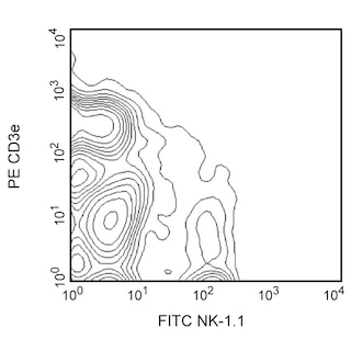
Detection of NK cells with monoclonal antibodies. Freshly isolated splenocytes from a C57BL/6 mouse were simultaneously incubated with FITC Mouse anti-Mouse NK-1.1 antibody (Cat. No. 553164), and PE Rat anti-Mouse CD49b antibody (clone DX5). Flow cytometry was performed on a BD FACScan™ Flow cytometer. (BD Biosciences, San Jose, CA)
.png)

BD Pharmingen™ PE Rat Anti-Mouse CD49b
.png)
Regulatory Status Legend
Any use of products other than the permitted use without the express written authorization of Becton, Dickinson and Company is strictly prohibited.
Preparation And Storage
Product Notices
- Since applications vary, each investigator should titrate the reagent to obtain optimal results.
- Please refer to www.bdbiosciences.com/us/s/resources for technical protocols.
- For fluorochrome spectra and suitable instrument settings, please refer to our Multicolor Flow Cytometry web page at www.bdbiosciences.com/colors.
- Caution: Sodium azide yields highly toxic hydrazoic acid under acidic conditions. Dilute azide compounds in running water before discarding to avoid accumulation of potentially explosive deposits in plumbing.
- An isotype control should be used at the same concentration as the antibody of interest.
Companion Products
.png?imwidth=320)

The rat anti-mouse CD49b monoclonal antibody (clone DX5) specifically binds to the integrin α2 chain (CD49b). CD49b is a 150 kDa transmembrane glycoprotein that non-covalently associates with CD29 (integrin β1) to form the integrin α2β1 complex known as VLA-2. The rat anti-mouse CD49b antibody (clone DX5) has been reported to identify the majority of NK cells and a small T-cell subpopulation in most mouse strains (e.g., A/J, AKR, BALB/c, C3H/HeJ, C57BL/6, C57BL/10, C57BR, C58, CBA/Ca, DBA/1, DBA/2, SJL, SWR, 129/J, but not NOD). The DX5 antibody also recognizes platelets that express high levels of CD49b. Multiparameter flow cytometric analysis has demonstrated that most lymphocytes which express NK-1.1 (NKR-P1B and NKR-P1C), as detectable by mouse anti-mouse NK-1.1 antibody (clone PK136), also express the DX5 antigen. Small DX5+ NK-1.1- and DX5- NK-1.1+ cell subsets are found, especially among the CD3-positive cell population. Some CD49b+ NK cells have been reported to gradually lose reactivity with the rat anti-mouse CD49b antibody (clone DX5) when cultured in the presence of recombinant human IL-2. The resulting DX5-negative cells have weakened cytotoxic activity when compared to the remaining DX5+ cells. This indicates that the DX5 antibody distinguishes functional subsets of NK cells. No activation or blocking activity of the rat anti-mouse antibody (clone DX5) has been observed. Staining of splenic NK cells with this antibody reportedly can be blocked by hamster anti-mouse CD49b antibody (clone HMα2).

Development References (3)
-
Arase H, Saito T, Phillips JH, Lanier LL. Cutting edge: the mouse NK cell-associated antigen recognized by DX5 monoclonal antibody is CD49b (alpha 2 integrin, very late antigen-2). J Immunol. 2001; 167(3):1141-1144. (Clone-specific: Blocking, Cytotoxicity, Flow cytometry). View Reference
-
Moore TA, von Freeden-Jeffry U, Murray R, Zlotnik A. Inhibition of gamma delta T cell development and early thymocyte maturation in IL-7 -/- mice. J Immunol. 1996; 157(6):2366-2373. (Biology). View Reference
-
Ortaldo JR, Winkler-Pickett R, Mason AT, Mason LH. The Ly-49 family: regulation of cytotoxicity and cytokine production in murine CD3+ cells. J Immunol. 1998; 160(1):1158-1165. (Biology). View Reference
Please refer to Support Documents for Quality Certificates
Global - Refer to manufacturer's instructions for use and related User Manuals and Technical data sheets before using this products as described
Comparisons, where applicable, are made against older BD Technology, manual methods or are general performance claims. Comparisons are not made against non-BD technologies, unless otherwise noted.
For Research Use Only. Not for use in diagnostic or therapeutic procedures.
Report a Site Issue
This form is intended to help us improve our website experience. For other support, please visit our Contact Us page.