Preparation And Storage
Recommended Assay Procedures
BD® CompBeads can be used as surrogates to assess fluorescence spillover (Compensation). When fluorochrome conjugated antibodies are bound to BD® CompBeads, they have spectral properties very similar to cells. However, for some fluorochromes there can be small differences in spectral emissions compared to cells, resulting in spillover values that differ when compared to biological controls. It is strongly recommended that when using a reagent for the first time, users compare the spillover on cells and BD CompBeads to ensure that BD® CompBeads are appropriate for your specific cellular application.
Product Notices
- Since applications vary, each investigator should titrate the reagent to obtain optimal results.
- An isotype control should be used at the same concentration as the antibody of interest.
- Caution: Sodium azide yields highly toxic hydrazoic acid under acidic conditions. Dilute azide compounds in running water before discarding to avoid accumulation of potentially explosive deposits in plumbing.
- For fluorochrome spectra and suitable instrument settings, please refer to our Multicolor Flow Cytometry web page at www.bdbiosciences.com/colors.
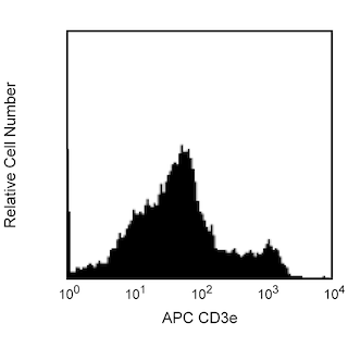
- This APC-conjugated reagent can be used in any flow cytometer equipped with a dye, HeNe, or red diode laser.
- Species cross-reactivity detected in product development may not have been confirmed on every format and/or application.
- Please refer to http://regdocs.bd.com to access safety data sheets (SDS).
- Please refer to www.bdbiosciences.com/us/s/resources for technical protocols.
Companion Products


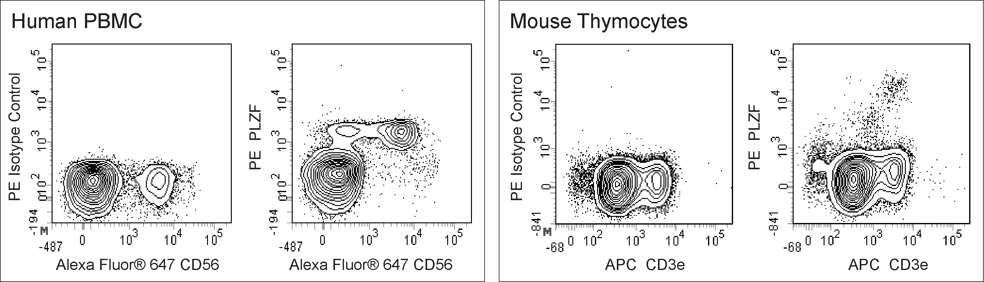
The R17-809 monoclonal antibody recognizes promyelocytic leukemia zinc finger protein (PLZF). PLZF is also known as zinc finger and BTB domain containing 16 (ZBTB16) and Zinc finger protein 145. PLZF is a member of the BTB/POZ-ZF family of transcription factors that includes Th-POK (Zbtb7b) which is involved in CD4+ T cell fate determination. PLZF serves multiple functions. In the immune system, PLZF is involved in the developmental regulation of innate immune lymphocytes including NKT cells, a subset of γδ T cells, and mucosal associated invariant T (MAIT) cells. The R17-809 hybridoma was generated from a mouse immunized with recombinant mouse PLZF protein. The R17-809 antibody crossreacts with both mouse and human PLZF.

Development References (3)
-
Alonzo ES, Gottschalk RA, Das J, et al. Development of promyelocytic zinc finger and ThPOK-expressing innate gamma delta T cells is controlled by strength of TCR signaling and Id3. J Immunol. 2010; 184(3):1268-1279. (Biology). View Reference
-
Alonzo ES, Sant'Angelo DB. Development of PLZF-expressing innate T cells. Curr Opin Immunol. 2011; 23(2):220-227. (Biology). View Reference
-
Savage AK, Constantinides MG, Han J, et al. The transcription factor PLZF directs the effector program of the NKT cell lineage. Immunity. 2008; 29(3):391-403. (Biology). View Reference
Please refer to Support Documents for Quality Certificates
Global - Refer to manufacturer's instructions for use and related User Manuals and Technical data sheets before using this products as described
Comparisons, where applicable, are made against older BD Technology, manual methods or are general performance claims. Comparisons are not made against non-BD technologies, unless otherwise noted.
For Research Use Only. Not for use in diagnostic or therapeutic procedures.