







Preparation And Storage


Recommended Assay Procedures
For optimal and reproducible results, BD Horizon Brilliant Stain Buffer should be used anytime two or more BD Horizon Brilliant dyes are used in the same experiment. Fluorescent dye interactions may cause staining artifacts which may affect data interpretation. The BD Horizon Brilliant Stain Buffer was designed to minimize these interactions. More information can be found in the Technical Data Sheet of the BD Horizon Brilliant Stain Buffer (Cat. No. 563794/566349) or the BD Horizon Brilliant Stain Buffer Plus (Cat. No. 566385).
Product Notices
- Since applications vary, each investigator should titrate the reagent to obtain optimal results.
- An isotype control should be used at the same concentration as the antibody of interest.
- Source of all serum proteins is from USDA inspected abattoirs located in the United States.
- Caution: Sodium azide yields highly toxic hydrazoic acid under acidic conditions. Dilute azide compounds in running water before discarding to avoid accumulation of potentially explosive deposits in plumbing.
- For fluorochrome spectra and suitable instrument settings, please refer to our Multicolor Flow Cytometry web page at www.bdbiosciences.com/colors.
- Alexa Fluor® is a registered trademark of Molecular Probes, Inc., Eugene, OR.
- BD Horizon Brilliant Violet 711 is covered by one or more of the following US patents: 8,110,673; 8,158,444; 8,227,187; 8,455,613; 8,575,303; 8,354,239.
- BD Horizon Brilliant Stain Buffer is covered by one or more of the following US patents: 8,110,673; 8,158,444; 8,575,303; 8,354,239.
- Cy is a trademark of GE Healthcare.
- Please refer to www.bdbiosciences.com/us/s/resources for technical protocols.
Companion Products
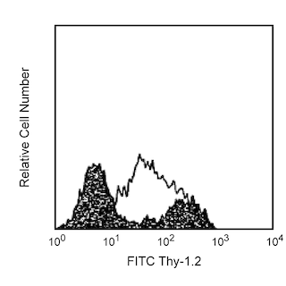

The C9B7W antibody monoclonal antibody specifically binds to an epitope in the D2 domain of CD223 (LAG3), the 70-kDa protein encoded by Lymphocyte-activation gene 3 (Lag3). A fusion protein consisting of the entire extracellular region of mouse LAG3 with mouse IgG1 was used as immunogen. CD223 is a type-I membrane protein with four extracellular Ig-like domains; it is structurally homologous to CD4; and, like CD4, it binds MHC class II molecules. However, unlike CD4, it is not expressed on resting human and mouse T lymphocytes. In the mouse, as previously described in the human, CD223 expression is upregulated on T lymphocytes (both CD4+ and CD8+) activated through the T-cell receptor (TCR) and on IL-2-activated NK (LAK) cells, and it is not detected on B cells, dendritic cells, or Phorbol 12-myristate 13-acetate (PMA)-stimulated splenocytes. Studies on human peripheral T lymphocytes suggest that CD223 associates with the TCR to downregulate TCR signaling. In contrast, in vivo and in vitro evaluations of vaccination protocols in mice suggest that CD223 promotes immune responses by activating antigen-presenting cells. Furthermore, NK cells of Lag3-/- mice display defects in their capacity to kill certain tumor cells. Mouse CD223 also has been demonstrated to contribute to the suppressor function of T regulatory cells and the C9B7W antibody has been shown to inhibit this function in vitro and in vivo. Therefore, CD223 appears to play complex roles in the regulation of immune responses. Although the C9B7W antibody is unable to block the binding of MHC class II-IgG2a fusion protein to CD223, it is able to block the CD223-mediated inhibition of IL-2 production by a T-cell hybridoma responding to antigen.
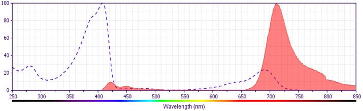
The antibody was conjugated to BD Horizon BV711 which is part of the BD Horizon Brilliant™ Violet family of dyes. This dye is a tandem fluorochrome of BD Horizon BV421 with an Ex Max of 405-nm and an acceptor dye with an Em Max at 711-nm. BD Horizon BV711 can be excited by the violet laser and detected in a filter used to detect Cy™5.5 / Alexa Fluor® 700-like dyes (eg, 712/20-nm filter). Due to the excitation and emission characteristics of the acceptor dye, there may be moderate spillover into the Alexa Fluor® 700 and PerCP-Cy5.5 detectors. However, the spillover can be corrected through compensation as with any other dye combination.
Development References (9)
-
Baixeras E, Huard B, Miossec C, et al. Characterization of the lymphocyte activation gene 3-encoded protein. A new ligand for human leukocyte antigen class II antigens. J Exp Med. 1992; 176(2):327-337. (Biology). View Reference
-
El Mir S, Triebel F. A soluble lymphocyte activation gene-3 molecule used as a vaccine adjuvant elicits greater humoral and cellular immune responses to both particulate and soluble antigens. J Immunol. 2000; 164(11):5583-5589. (Biology). View Reference
-
Hannier S, Tournier M, Bismuth G, Triebel F. CD3/TCR complex-associated lymphocyte activation gene-3 molecules inhibit CD3/TCR signaling. J Immunol. 1998; 161(8):4058-4065. (Biology). View Reference
-
Hannier S, Triebel F. The MHC class II ligand lymphocyte activation gene-3 is co-distributed with CD8 and CD3-TCR molecules after their engagement by mAb or peptide-MHC class I complexes. Int Immunol. 1999; 11(11):1745-1752. (Biology). View Reference
-
Huang CT, Workman CJ, Flies D, et al. Role of LAG-3 in regulatory T cells. Immunity. 2004; 21(4):503-513. (Clone-specific: Flow cytometry, Functional assay, Inhibition, In vivo exacerbation). View Reference
-
Huard B, Mastrangeli R, Prigent P, et al. Characterization of the major histocompatibility complex class II binding site on LAG-3 protein. Proc Natl Acad Sci U S A. 1997; 94(11):5744-5749. (Biology). View Reference
-
Miyazaki T, Dierich A, Benoist C, Mathis D. Independent modes of natural killing distinguished in mice lacking Lag3. Science. 1996; 272(5260):405-408. (Biology). View Reference
-
Prigent P, El Mir S, Dréano M, Triebel F. Lymphocyte activation gene-3 induces tumor regression and antitumor immune responses. Eur J Immunol. 1999; 29(12):3867-3876. (Biology). View Reference
-
Workman CJ, Rice DS, Dugger KJ, Kurschner C, Vignali DA. Phenotypic analysis of the murine CD4-related glycoprotein, CD223 (LAG-3). Eur J Immunol. 2002; 32(8):2255-2263. (Immunogen: Flow cytometry, Functional assay, Inhibition). View Reference
Please refer to Support Documents for Quality Certificates
Global - Refer to manufacturer's instructions for use and related User Manuals and Technical data sheets before using this products as described
Comparisons, where applicable, are made against older BD Technology, manual methods or are general performance claims. Comparisons are not made against non-BD technologies, unless otherwise noted.
For Research Use Only. Not for use in diagnostic or therapeutic procedures.
