







Preparation And Storage


Recommended Assay Procedures
BD™ CompBeads can be used as surrogates to assess fluorescence spillover (Compensation). When fluorochrome conjugated antibodies are bound to CompBeads, they have spectral properties very similar to cells. However, for some fluorochromes there can be small differences in spectral emissions compared to cells, resulting in spillover values that differ when compared to biological controls. It is strongly recommended that when using a reagent for the first time, users compare the spillover on cells and CompBead to ensure that BD Comp beads are appropriate for your specific cellular application.
For optimal and reproducible results, BD Horizon Brilliant Stain Buffer should be used anytime two or more BD Horizon Brilliant dyes are used in the same experiment. Fluorescent dye interactions may cause staining artifacts which may affect data interpretation. The BD Horizon Brilliant Stain Buffer was designed to minimize these interactions. More information can be found in the Technical Data Sheet of the BD Horizon Brilliant Stain Buffer (Cat. No. 563794/566349) or the BD Horizon Brilliant Stain Buffer Plus (Cat. No. 566385).
Product Notices
- Since applications vary, each investigator should titrate the reagent to obtain optimal results.
- An isotype control should be used at the same concentration as the antibody of interest.
- Caution: Sodium azide yields highly toxic hydrazoic acid under acidic conditions. Dilute azide compounds in running water before discarding to avoid accumulation of potentially explosive deposits in plumbing.
- Source of all serum proteins is from USDA inspected abattoirs located in the United States.
- Alexa Fluor® is a registered trademark of Molecular Probes, Inc., Eugene, OR.
- For fluorochrome spectra and suitable instrument settings, please refer to our Multicolor Flow Cytometry web page at www.bdbiosciences.com/colors.
- BD Horizon Brilliant Violet 650 is covered by one or more of the following US patents: 8,110,673; 8,158,444; 8,227,187; 8,455,613; 8,575,303; 8,354,239.
- BD Horizon Brilliant Stain Buffer is covered by one or more of the following US patents: 8,110,673; 8,158,444; 8,575,303; 8,354,239.
- Please refer to http://regdocs.bd.com to access safety data sheets (SDS).
- Please refer to www.bdbiosciences.com/us/s/resources for technical protocols.
Companion Products
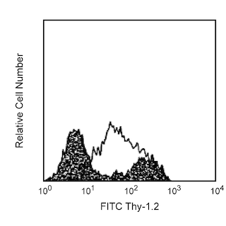



The 2G8 monoclonal antibody specifically binds to the mouse C-X-C Chemokine Receptor type 5, CXCR5. CXCR5 is also known as CD185, BLR1, NLR and MDR15. CXCR5 is a seven-transmembrane, G-protein-coupled receptor that is specific for the CXC chemokine, CXCL13/BLC/BCA-1. The expression of CXCR5 has been detected in spleen, lymph nodes, tonsils, brain, bone marrow, T cells, B cells, cerebrum, cerebellum, hippcampus and pituitary. In mouse spleen, CXCR5 was strictly expressed by mature B cells and a small subset of T lymphocytes. CXCR5 plays a role in directing the migration of B and T cells to B cell follicles with the spleen and certain other lymphoid tissues. The immunogen used to generate 2G8 hybridoma was a recombinant protein containing N-terminal amino acids of mouse CXCR5 (GST-NmBLR1).
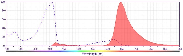
The antibody was conjugated to BD Horizon BV650 which is part of the BD Horizon Brilliant™ Violet family of dyes. This dye is a tandem fluorochrome of BD Horizon BV421 with an Ex Max of 405-nm and an acceptor dye with an Em Max at 650-nm. BD Horizon BV650 can be excited by the violet laser and detected in a filter used to detect APC-like dyes (eg, 660/20-nm filter). Due to the excitation and emission characteristics of the acceptor dye, there will be spillover into the APC and Alexa Fluor® 700 detectors. However, the spillover can be corrected through compensation as with any other dye combination.
Development References (6)
-
Barella L, Loetscher M, Tobler A, Baggiolini M, Moser B. Sequence variation of a novel heptahelical leucocyte receptor through alternative transcript formation. Biochem J. 1995; 309(3):773-779. (Biology). View Reference
-
Dobner T, Wolf I, Emrich T, Lipp M. Differentiation-specific expression of a novel G protein-coupled receptor from Burkitt's lymphoma. Eur J Immunol. 1992; 22(11):2795-2799. (Biology). View Reference
-
Forster R, Mattis AE, Kremmer E, Wolf E, Brem G, Lipp M. A putative chemokine receptor, BLR1, directs B cell migration to defined lymphoid organs and specific anatomic compartments of the spleen. Cell. 1996; 87(6):1037-1047. (Immunogen: ELISA, Flow cytometry, Immunofluorescence, Immunohistochemistry). View Reference
-
Gunn MD, Ngo VN, Ansel KM, Ekland EH, Cyster JG, Williams LT. A B-cell-homing chemokine made in lymphoid follicles activates Burkitt's lymphoma receptor-1. Nature. 1998; 391(6669):799-803. (Biology). View Reference
-
Kaiser E, Forster R, Wolf I, Ebensperger C, Kuehl WM, Lipp M. The G protein-coupled receptor BLR1 is involved in murine B cell differentiation and is also expressed in neuronal tissues. Eur J Immunol. 1993; 23(10):2532-2539. (Biology). View Reference
-
Kouba M, Vanetti M, Wang X, Schafer M, Hollt V. Cloning of a novel putative G-protein-coupled receptor (NLR) which is expressed in neuronal and lymphatic tissue. FEBS Lett. 1993; 321(2-3):173-178. (Biology). View Reference
Please refer to Support Documents for Quality Certificates
Global - Refer to manufacturer's instructions for use and related User Manuals and Technical data sheets before using this products as described
Comparisons, where applicable, are made against older BD Technology, manual methods or are general performance claims. Comparisons are not made against non-BD technologies, unless otherwise noted.
For Research Use Only. Not for use in diagnostic or therapeutic procedures.
