Old Browser
This page has been recently translated and is available in French now.
Looks like you're visiting us from {countryName}.
Would you like to stay on the current country site or be switched to your country?


Multiparameter flow cytometric analysis of CD45 expression on Rhesus macaque peripheral blood leucocytes. Rhesus macaque whole blood was stained with either BD Horizon™ BV480 Mouse IgG1, κ Isotype Control (Cat. No. 565652; Left Plot) or BD Horizon BV480 Mouse Anti-NHP CD45 antibody (Cat. No. 566145/566152; Right Plot). The erythrocytes were lysed with BD FACS™ Lysing Solution (Cat. No. 349202). Two parameter flow cytometric contour plots showing the correlated expression of CD45 (or Ig Isotype control staining) versus side-light scatter (SSC-A) signals were derived from events with the forward light scatter characteristics of intact leucocyte populations. Flow cytometric analysis was performed using a BD LSRFortessa™ Cell Analyzer System.


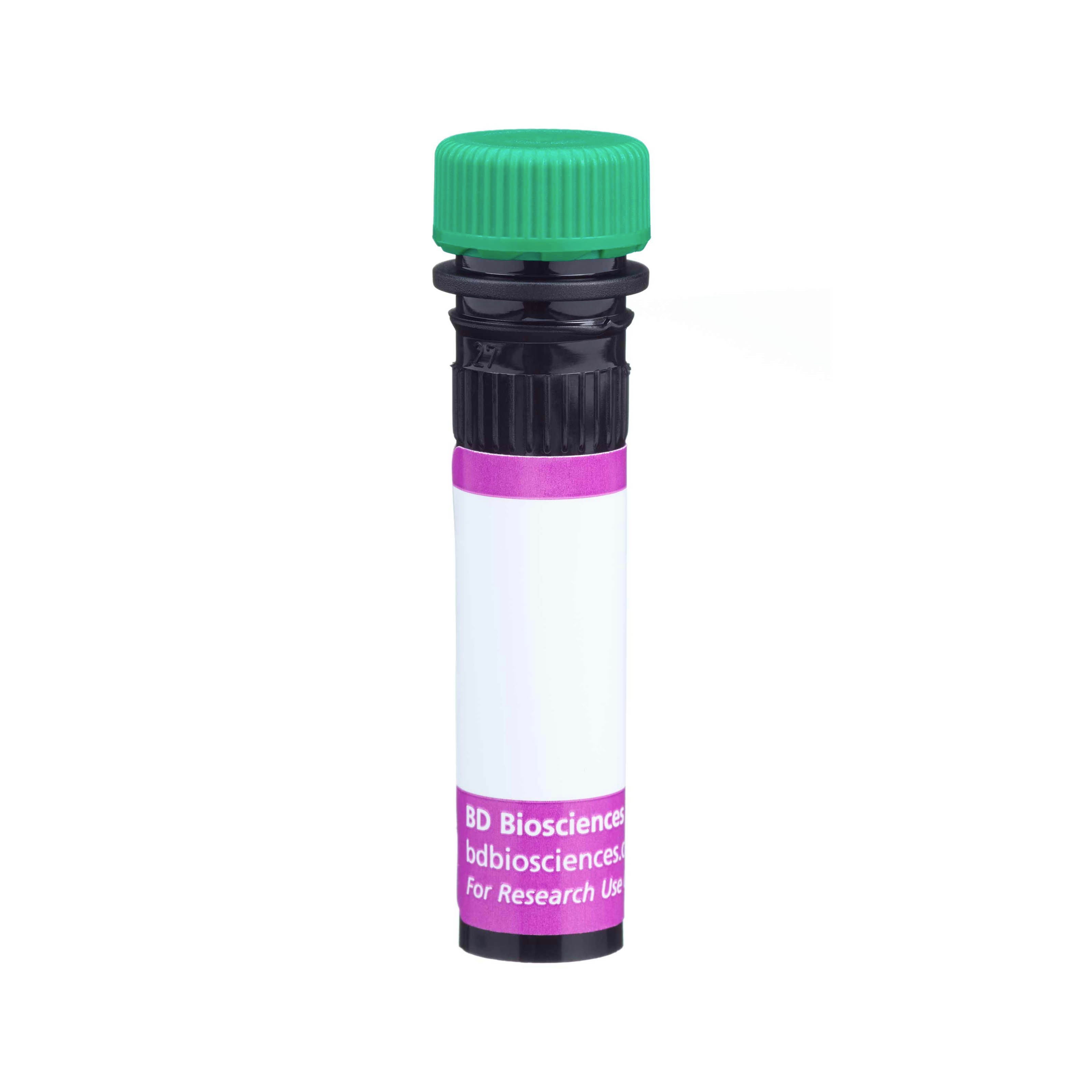
BD Horizon™ BV480 Mouse Anti-NHP CD45

Regulatory Status Legend
Any use of products other than the permitted use without the express written authorization of Becton, Dickinson and Company is strictly prohibited.
Preparation And Storage
Recommended Assay Procedures
For optimal and reproducible results, BD Horizon Brilliant™ Stain Buffer should be used anytime BD Horizon Brilliant™ dyes are used in a multicolor flow cytometry panel. Fluorescent dye interactions may cause staining artifacts which may affect data interpretation. The BD Horizon Brilliant Stain Buffer was designed to minimize these interactions. When BD Horizon Brilliant Stain Buffer is used in in the multicolor panel, it should also be used in the corresponding compensation controls for all dyes to achieve the most accurate compensation. For the most accurate compensation, compensation controls created with either cells or beads should be exposed to BD Horizon Brilliant Stain Buffer for the same length of time as the corresponding multicolor panel. More information can be found in the Technical Data Sheet of the BD Horizon Brilliant Stain Buffer (Cat. No. 563794/566349) or the BD Horizon Brilliant Stain Buffer Plus (Cat. No. 566385).
Product Notices
- This reagent has been pre-diluted for use at the recommended Volume per Test. We typically use 1 × 10^6 cells in a 100-µl experimental sample (a test).
- An isotype control should be used at the same concentration as the antibody of interest.
- Source of all serum proteins is from USDA inspected abattoirs located in the United States.
- Caution: Sodium azide yields highly toxic hydrazoic acid under acidic conditions. Dilute azide compounds in running water before discarding to avoid accumulation of potentially explosive deposits in plumbing.
- For fluorochrome spectra and suitable instrument settings, please refer to our Multicolor Flow Cytometry web page at www.bdbiosciences.com/colors.
- BD Horizon Brilliant Violet 480 is covered by one or more of the following US patents: 8,575,303; 8,354,239.
- BD Horizon Brilliant Stain Buffer is covered by one or more of the following US patents: 8,110,673; 8,158,444; 8,575,303; 8,354,239.
- Species cross-reactivity detected in product development may not have been confirmed on every format and/or application.
- Please refer to www.bdbiosciences.com/us/s/resources for technical protocols.
Companion Products


D058-1283 is a CD45 monoclonal antibody specific for non-human primate leucocytes. It was developed using Rhesus peripheral whole blood as the immunogen. It does not cross-react with human leucocytes. This antibody reacts with baboon, Rhesus and Cynomolgus Macaque leucocytes in a similar pattern to CD45 binding to leukocyte common antigen (LCA) on human cells. Immunophenotypic analysis shows that D058-1283 binds to lymphocytes, monocytes and granulocytes of non-human primate blood samples. This antibody is able to block the binding of monoclonal antibody TÜ116; a reported anti-human CD45 antibody that cross-reacts with nonhuman primate leucocytes. In Western blot analysis, the D058-1283 antibody identifies a 180-200 kDa band.

Development References (4)
-
Brown KN, Trichel A, Barratt-Boyes SM. Parallel loss of myeloid and plasmacytoid dendritic cells from blood and lymphoid tissue in simian AIDS. J Immunol. 2007; 178(11):6958-6967. (Clone-specific: Flow cytometry). View Reference
-
Drouet M, Mayol JF, Norol F, et al. Lack of evidence of sustained hematopoietic reconstitution after transplantation of unmanipulated adult liver stem cells in monkeys. Haematologica. 2007; 92(2):248-251. (Clone-specific: Flow cytometry). View Reference
-
Reeves RK, Evans TI, Gillis J, et al. Quantification of mucosal mononuclear cells in tissues with a fluorescent bead-based polychromatic flow cytometry assay. J Immunol Methods. 2011; 367(1-2):95-98. (Clone-specific: Flow cytometry). View Reference
-
Reimann KA, Waite BC, Lee-Parritz DE, et al. Use of human leukocyte-specific monoclonal antibodies for clinically immunophenotyping lymphocytes of rhesus monkeys. Cytometry. 1994; 17(1):102-108. (Biology). View Reference
Please refer to Support Documents for Quality Certificates
Global - Refer to manufacturer's instructions for use and related User Manuals and Technical data sheets before using this products as described
Comparisons, where applicable, are made against older BD Technology, manual methods or are general performance claims. Comparisons are not made against non-BD technologies, unless otherwise noted.
For Research Use Only. Not for use in diagnostic or therapeutic procedures.
Report a Site Issue
This form is intended to help us improve our website experience. For other support, please visit our Contact Us page.