
Preparation And Storage
Product Notices
- Since applications vary, each investigator should titrate the reagent to obtain optimal results.
- An isotype control should be used at the same concentration as the antibody of interest.
- Caution: Sodium azide yields highly toxic hydrazoic acid under acidic conditions. Dilute azide compounds in running water before discarding to avoid accumulation of potentially explosive deposits in plumbing.
- The Alexa Fluor®, Pacific Blue™, and Cascade Blue® dye antibody conjugates in this product are sold under license from Molecular Probes, Inc. for research use only, excluding use in combination with microarrays, or as analyte specific reagents. The Alexa Fluor® dyes (except for Alexa Fluor® 430), Pacific Blue™ dye, and Cascade Blue® dye are covered by pending and issued patents.
- Alexa Fluor® is a registered trademark of Molecular Probes, Inc., Eugene, OR.
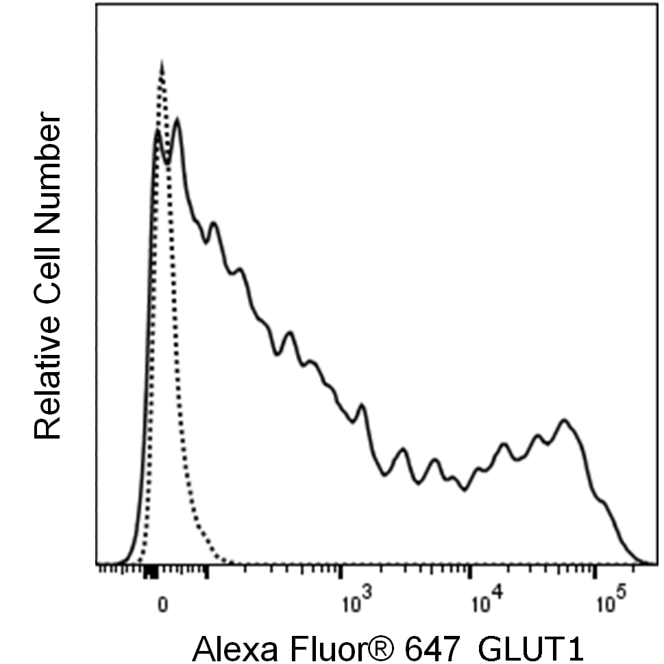
- Alexa Fluor® 647 fluorochrome emission is collected at the same instrument settings as for allophycocyanin (APC).
- For fluorochrome spectra and suitable instrument settings, please refer to our Multicolor Flow Cytometry web page at www.bdbiosciences.com/colors.
- Please refer to www.bdbiosciences.com/us/s/resources for technical protocols.
Companion Products


The monoclonal antibody 202915 specifically recognizes Glucose transporter 1 (GLUT1) that is encoded by SLC2A1 (Solute carrier family 2, facilitated glucose transporter member 1). GLUT1 serves as a transporter for glucose and other aldose sugars and oxidized ascorbic acid (vitamin C) into cells. GLUT1 is also known as the Receptor for HTLV-1 and HTLV-2 (HTLVR). Structural studies suggest that GLUT1 is a twelve-pass transmembrane glycoprotein (12TM) with cytoplasmic N- and C-termini. GLUT1 plays an important role in the glycolytic pathway by transporting glucose across the plasma membranes of cells. GLUT1 is differentially expressed on a wide variety of normal cells including erythrocytes, blood-brain barrier endothelial cells, astrocytes, adipocytes, cardiomyocytes, lymphocytes and other leucocytes, as well as by tumor cells and cell lines.
Development References (5)
-
Basu S, Hubbard B, Shevach EM. Foxp3-mediated inhibition of Akt inhibits Glut1 (glucose transporter 1) expression in human T regulatory cells. J Leukoc Biol. 2015; 97(2):279-283. (Biology). View Reference
-
Carruthers A, DeZutter J, Ganguly A, Devaskar S. Will the original glucose transporter isoform please stand up!. Amer J Physiol Endocrinol Metab. 2009; 297(4):E836-848. (Biology). View Reference
-
Chan O, Burke JD, Gao DF, Fish EN. The chemokine CCL5 regulates glucose uptake and AMP kinase signaling in activated T cells to facilitate chemotaxis. J Biol Chem. 2012; 287(35):29406-29416. (Biology). View Reference
-
Kinet S, Swainson L, Lavanya M, et al. Isolated receptor binding domains of HTLV-1 and HTLV-2 envelopes bind Glut-1 on activated CD4+ and CD8+ T cells. Retrovirology. 2007; 4:31. (Biology). View Reference
-
Takenouchi N, Jones KS, Lisinski I, et al. GLUT1 is not the primary binding receptor but is associated with cell-to-cell transmission of human T-cell leukemia virus type 1. J Virol. 2007; 81(3):1506-1510. (Biology). View Reference
Please refer to Support Documents for Quality Certificates
Global - Refer to manufacturer's instructions for use and related User Manuals and Technical data sheets before using this products as described
Comparisons, where applicable, are made against older BD Technology, manual methods or are general performance claims. Comparisons are not made against non-BD technologies, unless otherwise noted.
For Research Use Only. Not for use in diagnostic or therapeutic procedures.