Skip to main content Skip to navigation
DRAQ7™

BD Pharmingen™ DRAQ7™

(RUO)
DRAQ7™
Panel 1. Two-color flow cytometric analysis of Jurkat cell viability. Jurkat cells were treated with 5 µM Camptothecin (Right Plot) or DMSO vehicle (Left Plot) overnight. Cells were resuspended in Annexin V Binding Buffer (Cat. No. 556454) and stained with FITC Annexin V (Cat. No. 556419) and 1.25 µM DRAQ7™ (Cat. No. 564904). Camptothecin-treated cells show an increased frequency of apoptotic (Annexin V+ DRAQ7™-) and dead (Annexin V+ DRAQ7™+) cells. Analysis was performed using a BD LSRFortessa™ Cell Analyzer System.   Panel 2. Flow cytometric analysis of HeLa cell DNA content. Cultured HeLa cells in log phase growth were harvested using Gibco® Cell Dissociation Buffer (Life Technologies), fixed, and permeabilized using the BD Pharmingen™ Transcription Factor Buffer Set (Cat. No. 562574/562725). Cells were resuspended in DPBS with 20 μM DRAQ7™ and analyzed using a low BD LSRFortessa™ cytometer flow rate. Histograms were deconvoluted by FlowJo™ software into G0/G1, S, and G2/M populations. Panel 3. Immunoflourescent staining of Alkaline Phosphatase on human embryonic stem (ES) cells. H9 human ES cells (WiCell, Madison, WI) passage 44 were cultured on mTeSR™1 medium (StemCell Technologies) and fixed with BD Cytofix™ fixation buffer (Cat. No. 554655). The fixed cells were stained with Alexa Fluor® 488 Mouse anti-Human Alkaline Phosphatase monoclonal antibody (pseudo-colored green, Cat. No. 561495). And 5 μM DRAQ7™ (pseudo-colored red) was used as a nuclear counterstain. The images were captured on a BD Pathway™ 435 Cell Analyzer and merged using BD Attovision™ Software. DRAQ7™ was also tested in mouse (data not shown).
Product Details
Down Arrow Up Arrow


Description

DRAQ7™ (Deep Red Anthraquinone 7) is a far-red fluorescent DNA dye. DRAQ7™ is cell impermeable and may be used to stain nucleic acids in fixed cells for cell cycle analysis by DNA content, nuclear visualization, or discrimination of nucleated cells from debris or enucleated cells. Because DRAQ7 is impermeable to intact cells, it may also be used as a viability dye. DRAQ7™ has an excitation wavelength maxima of 599/644 nm, but can also be suboptimally excited by the 488 nm wavelength laser. Its emission wavelength maximum is 678 nm, or 694 nm when intercalated with double-stranded DNA.


Preparation And Storage

Store undiluted at 4°C and protected from prolonged exposure to light. Do not freeze.

Recommended Assay Procedures

Staining of Live Cells for Viability Analysis by Flow Cytometry

1.        Obtain a single cell suspension.

2.        Resuspend cells in 1× Dulbecco's Phosphate Buffered Saline (DPBS) or other azide-free buffer containing 1-3 μM DRAQ7™.

a.        The optimal concentration of DRAQ7™ for viability analysis may vary by cell type. We recommend titrating the reagent for your cell type of interest in early experiments.

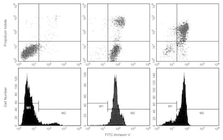
b.        Additionally, apoptotic cells may stain with variable amounts of DRAQ7™. We recommend co-staining with, eg, BD Pharmingen™ FITC  Annexin V (Cat. No. 556419) if further analysis of apoptotic cells is desired.

3.        Incubate 5 minutes at room temperature. No wash is necessary prior to analysis.

4.        Proceed to analysis by flow cytometry.

Staining of Fixed Cells for DNA Content Analysis by Flow Cytometry

1.        Obtain a single cell suspension.

2.        Treat cells on ice for 30 minutes with 70-80% ice-cold ethanol.

a.        Ethanol fixation typically provides the most resolved histograms. However, this reagent has also been successfully used for DNA content analysis with the BD Pharmingen™  Transcription Factor Buffer Set (Cat. No. 562574/562725) or BD Cytofix™ Fixation Buffer (Cat. No. 554655) and BD Phosflow™ Perm III (Cat. No. 558050) protocol.

3.        Wash cells once with BD Pharmingen™ Stain Buffer (FBS) (Cat. No. 554656).

4.        Dilute DRAQ7™ to 20 μM in 1× DPBS or other azide-free buffer immediately prior to use.

5.        Stain cells for 5-15 minutes at a cell density of 0.5E6 cells/mL or less. No further wash is necessary prior to analysis.

a.        The optimal cell density and concentration of DRAQ7™ for DNA content analysis may vary by cell type. Assay conditions should be optimized in early experiments for best results.

6.        Proceed to analysis by flow cytometry.

Immunofluorescent Staining of Fixed Cells for Nuclear Visualization

1.        Fix and permeabilize cells as desired.

2.        Dilute DRAQ7™ solution to 5-20 μM in 1× DPBS or other azide-free buffer immediately prior to use.

3.        Add DRAQ7™ solution to each sample at least 5 minutes before analysis.

4.        Proceed to imaging. We recommend using a 715LP or longer wavelength filter, though the dye is well-detected in filters typically used to detect Alexa Fluor® 647 (eg, 660/20 or 692/40). Note that dsDNA-bound dye will fluoresce brightly in the nucleus and unbound dye may fluoresce dimly in the cytoplasm, allowing segmentation of the cytoplasmic and nuclear compartments.

Note: This reagent has been developed and certified for the Bioimaging application. However, a routine Bioimaging test is not performed on every lot.

Warning:  DRAQ7™ contains < 1% 1,5-BIS{[2-(DIMETHYLAMINO)ETHYL]AMINO}-4,8-DIHYDROXYANTHRACENE-9,10-DIONE

Hazard statements

Causes skin irritation.

Causes serious eye irritation.

May cause respiratory irritation.

Precautionary statements

Wear protective gloves/protective clothing/eye protection/face protection.

Wash thoroughly after handling.

IF IN EYES: Rinse cautiously with water for several minutes. Remove contact lenses, if present and easy to do.

Continue rinsing. If eye irritation persists: Get medical advice/attention.

Take off contaminated clothing and wash before reuse.

Call a POISON CENTER or doctor/physician if you feel unwell.

If skin irritation occurs: Get medical advice/attention. IF ON SKIN: Wash with plenty of water

Avoid breathing dust/fume/gas/mist/vapors/spray.

Use only outdoors or in a well-ventilated area.

IF INHALED: Remove person to fresh air and keep comfortable for breathing.  

Store in a well-ventilated place. Keep container tightly closed.  Store locked up.

Dispose of contents/container to an appropriate treatment and disposal facility in accordance with applicable laws and regulations, and product characteristics at time of disposal.

Product Notices

  1. DRAQ7™ is a registered trademark of BioStatus Ltd.
  2. FlowJo is a trademark of Tree Star Inc.
  3. Alexa Fluor® is a registered trademark of Molecular Probes, Inc., Eugene, OR.
  4. mTESR™1 is a trademark of StemCell Technologies.
  5. This antibody has been developed and certified for the bioimaging application. However, a routine bioimaging test is not performed on every lot. Researchers are encouraged to titrate the reagent for optimal performance.
  6. Please refer to www.bdbiosciences.com/us/s/resources for technical protocols.
564904 Rev. 2
Citations & References
Down Arrow Up Arrow

Development References (5)

  1. Akagi J, Kordon M, Zhao H, et al. Real-time cell viability assays using a new anthracycline derivative DRAQ7®. Cytometry A. 2013; 83(2):227-234. (Methodology). View Reference
  2. Edward R. Red/far-red fluorescing DNA-specific anthraquinones for nucl:cyto segmentation and viability reporting in cell-based assays. Methods Enzymol. 2012; 505:23-45. (Methodology). View Reference
  3. Smith PJ, Blunt N, Wiltshire M, et al. Characteristics of a novel deep red/infrared fluorescent cell-permeant DNA probe, DRAQ5, in intact human cells analyzed by flow cytometry, confocal and multiphoton microscopy. Cytometry. 2000; 40(4):280-291. (Methodology). View Reference
  4. Smith PJ, Wiltshire M, Davies S, et al. A novel cell permeant and far red-fluorescing DNA probe, DRAQ5, for blood cell discrimination by flow cytometry. J Immunol Methods. 1999; 229(1):131-139. (Methodology). View Reference
  5. Smith PJ, Wiltshire M, Errington RJ. DRAQ5 Labeling of Nuclear DNA in Live and Fixed Cells. Curr Protoc Cytom. 7(7.25)(Methodology). View Reference
View All (5) View Less
564904 Rev. 2

Please refer to Support Documents for Quality Certificates

Global - Refer to manufacturer's instructions for use and related User Manuals and Technical data sheets before using this products as described

Comparisons, where applicable, are made against older BD Technology, manual methods or are general performance claims.  Comparisons are not made against non-BD technologies, unless otherwise noted.

For Research Use Only. Not for use in diagnostic or therapeutic procedures.