Preparation And Storage
The antibody reagent is stable until the expiration date shown on the label when stored at 2° to 8°C. Do not use after the expiration date. Do not freeze the reagent or expose it to direct light during storage or incubation with cells. Keep the outside of the reagent vial dry.
Do not use the reagent if you observe any change in appearance. Precipitation or discoloration indicates instability or deterioration.
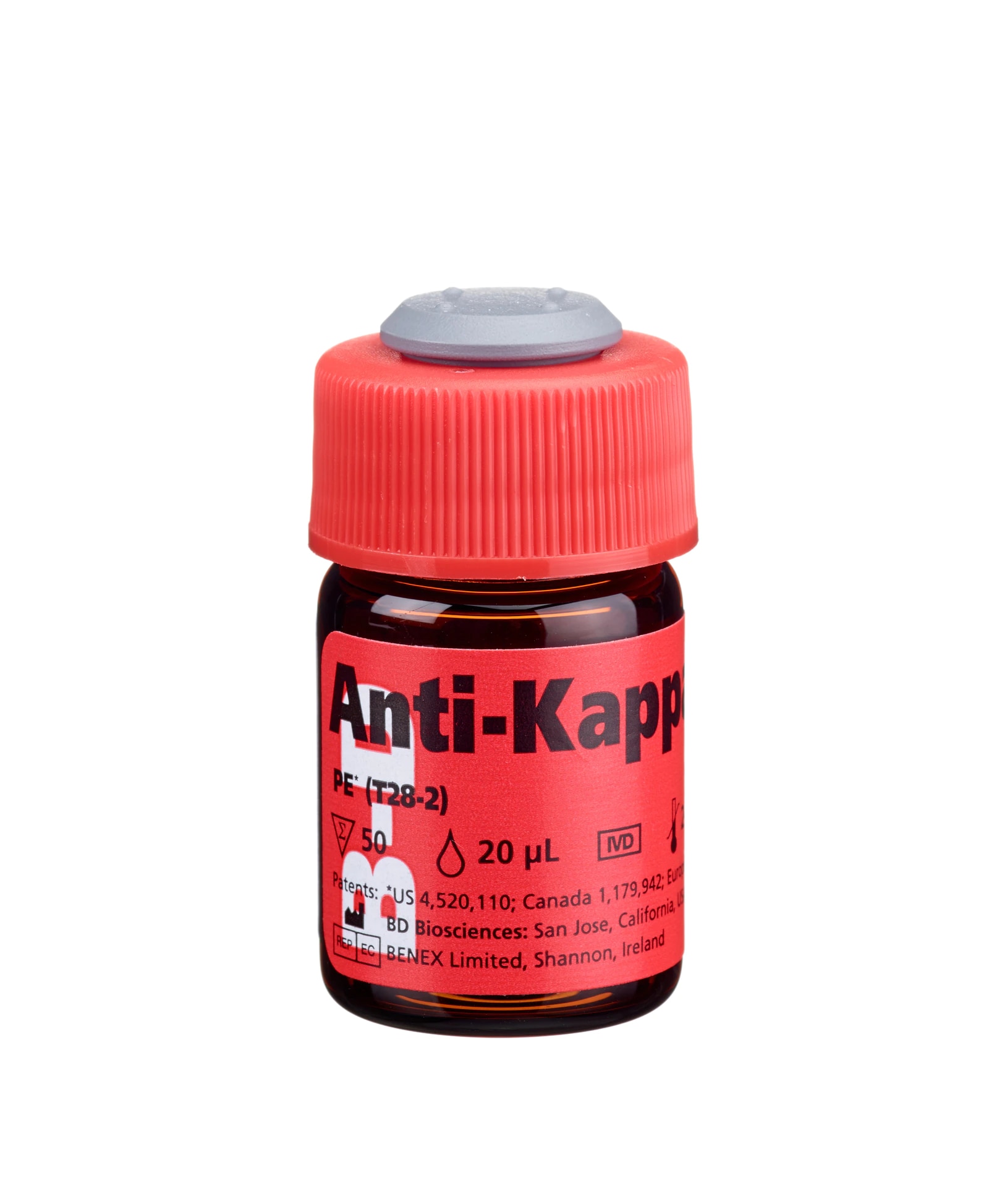
Anti-Kappa is intended for in vitro diagnostic use in the identification of cells expressing kappa light chains, using a BD FACS™ brand flow cytometer. The flow cytometer must be equipped to detect light scatter and the appropriate fluorescence, and be equipped with appropriate analysis software for data acquisition and analysis. See your instrument user’s guide for instructions.

Development References (25)
-
Agrawal YP, Hamalainen E, Mahlamaki EK, et al. Comparison of poly- and monoclonal antibodies for determination of B-cell clonal excess in an routine clinical laboratory. Eur J Haematol. 1992; 48:49-55. (Biology).
-
Ault KA. Flow cytometric evaluation of normal and neoplastic B cells. In: Rose NR, Friedman H, Fahey JL. Rose NR, Friedman H, Fahey JL, ed. Manual of Clinical Laboratory Immunology. 3rd ed.. Washington, DC: American Society for Microbiology; 1986:247-253. View Reference
-
Berliner N, Ault KA, Martin P, Weinberg DS. Detection of clonal excess in lymphoproliferative disease by κ/λ analysis: correlation with immunoglobulin gene DNA rearrangement. Blood. 1986; 67:80-85. (Biology).
-
Braylan RC, Benson NA, Iturraspe J. Analysis of lymphomas by flow cytometry: current and emerging strategies. Ann NY Acad Sci. 1993; 677:364-378. (Biology).
-
Centers for Disease Control. Update: universal precautions for prevention of transmission of human immunodeficiency virus, hepatitis B virus, and other bloodborne pathogens in healthcare settings. MMWR. 1988; 37:377-388. (Biology).
-
Chizuka A, Kanda Y, Nannya Y, et al. The diagnostic value of κ/λ ratios determined by flow cytometric analysis of biopsy specimens in B-cell lymphoma. Clin Lab Haematol. 2002; 24:33-36. (Biology).
-
Clinical Applications of Flow Cytometry: Quality Assurance and Immunophenotyping of Lymphocytes: Approved Guideline. H42-A2. 2007. (Biology).
-
Clinical and Laboratory Standards Institute. 2005. (Biology).
-
Consensus protocol for the flow cytometric immunophenotyping of hematopoietic malignancies. Rothe G, Schmitz G. Leukemia. 1996; 10:877-895. (Biology).
-
Foon KA, Todd RF. Immunologic classification of leukemia and lymphoma.. Blood. 1986; 68(1):1-31. (Biology). View Reference
-
Fukushima PI, Nguyen PK, O'Grady P, Stetler-Stevenson M. Flow cytometric analysis of kappa and lambda light chain expression in evaluation of specimens for B-cell neoplasia.. Cytometry. 1996; 26(4):243-52. (Biology). View Reference
-
Harris NL, Data RE. The distribution of neoplastic and normal B-lymphoid cells in nodular lymphomas: use of an immunoperoxidase technique on frozen sections.. Hum Pathol. 1982; 13(7):610-7. (Biology). View Reference
-
Jackson AL, Warner NL. Rose NR, Friedman H, Fahey JL, ed. Manual of Clincial Laboratory Immunology, Third Edition. Washington DC: American Society for Microbiology; 1986:226-235.
-
Maiese RL, Segal GH, Iturraspe JA, Braylan RC. The cell surface antigen and DNA content distribution of lymph nodes with reactive hyperplasia. Mod Pathol. 1995; 8:536-543. (Biology).
-
Meis JM, Osborne BM, Butler JJ. A comparative marker study of large cell lymphoma, Hodgkin's disease, and true histiocytic lymphoma in paraffin-embedded tissue.. Am J Clin Pathol. 1986; 86(5):591-9. (Biology). View Reference
-
Oertel J, Lipski S, Lobeck H, Huhn D. Detection of light chain restriction in chronic B-lymphoid leukaemia and B-non-Hodgkin's lymphoma. Clin Lab Haematol. 1991; 13:33-40. (Biology).
-
Picker LJ, Weiss LM, Medeiros LJ, Wood GS, Warnke RA. Immunophenotypic criteria for the diagnosis of non-Hodgkin's lymphoma.. Am J Pathol. 1987; 128(1):181-201. (Biology). View Reference
-
Siebert JD, Mulvaney DA, Vukov AM, Knost JA, King DE, Craig FE. Utility of flow cytometry in subtyping composite and sequential lymphoma. J Clin Lab Anal. 1999; 13:199-204. (Biology).
-
Smith BR, Weinberg DS, Robert NJ, et al. Circulating monoclonal B lymphocytes in non-Hodgkin's lymphoma.. N Engl J Med. 1984; 311(23):1476-81. (Biology). View Reference
-
Stelzer GT, Marti G, Hurley A, McCoy PJ, Lovett EJ, Schwartz A. US-Canadian consensus recommendations on the immunophenotypic analysis of hematologic neoplasia by flow cytometry: standardization and validation of laboratory procedures. Cytometry. 1997; 30:214-230. (Biology).
-
Stetler-Stevenson M, Braylan RC. Flow cytometric analysis of lymphomas and lymphoproliferative disorders.. Semin Hematol. 2001; 38(2):111-23. (Biology). View Reference
-
Tubbs RR, Sheibani K, Weiss RA, Sebek BA, Deodhar SD. Tissue immunomicroscopic evaluation of monoclonality of B-cell lymphomas: comparison with cell suspension studies.. Am J Clin Pathol. 1981; 76(1):24-8. (Biology). View Reference
-
Tworek JA, Singleton TP, Schnitzer B, Hsi ED, Ross CW. Flow cytometric and immunohistochemical analysis of small lymphocytic lymphoma, mantle cell lymphoma, and plasmacytoid small lymphocytic lymphoma. Am J Clin Pathol. 1998; 110:582-589. (Biology).
-
Têtu B, Manning JT, Ordóñez NG. Comparison of monoclonal and polyclonal antibodies directed against immunoglobulin light and heavy chains in non-Hodgkin's lymphoma.. Am J Clin Pathol. 1986; 85(1):25-31. (Biology). View Reference
-
Weinberg DS, Pinkus GS, Ault KA. Cytofluorometric detection of B cell clonal excess: a new approach to the diagnosis of B cell lymphoma.. Blood. 1984; 63(5):1080-7. (Biology). View Reference
Please refer to Support Documents for Quality Certificates
Global - Refer to manufacturer's instructions for use and related User Manuals and Technical data sheets before using this products as described
Comparisons, where applicable, are made against older BD Technology, manual methods or are general performance claims. Comparisons are not made against non-BD technologies, unless otherwise noted.
For In Vitro Diagnostic Use.
23-22942-00
![]() Documents are subject to revision without notice. Please verify you have the correct revision of the document, and always refer back to BD's eIFU website for the latest and most up to date information.
Documents are subject to revision without notice. Please verify you have the correct revision of the document, and always refer back to BD's eIFU website for the latest and most up to date information.