


Protocols
Featured Protocols
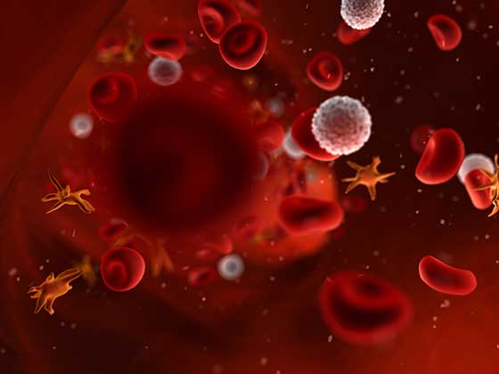
RBC/Platelet Protocols
Get protocols for surface staining and intracellular staining of human red blood cells and indirect immunofluorescence of human platelets. Find the procedure for platelet activation, staining and analysis.
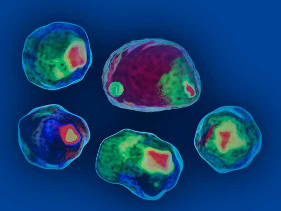
Flow Cytometry Protocols
Explore protocols for sample preparation of mouse and rat leucocytes, indirect staining of mononuclear cells, reducing nonspecific staining with Fc Block, immune cell activation.
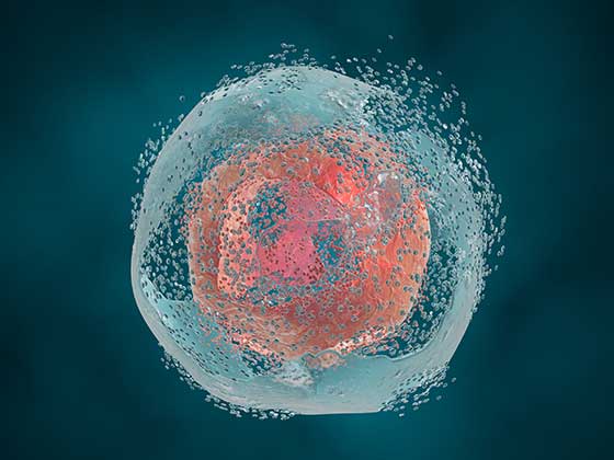
Apoptosis Protocols
Find protocols for induction of apoptosis using anti-Fas antibodies or by using various inhibitors. Get step-by-step protocol for Annexin V staining or for studying DNA fragmentation using the BD® Apo-BrdU™ kit.
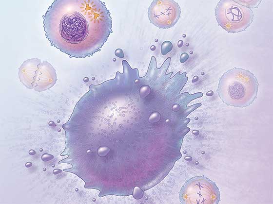
Cell Biology Protocols
Explore protocols for immunoprecipitation with antibody:agarose or anti-phosphotyrosine:biotin conjugates. Western blotting protocols with monoclonal, polyclonal, HRP- and biotin-conjugated antibodies are also available.