





BD Pharmingen™ Purified Mouse Anti-Human CD140a
Clone αR1 (also known as Alpha-R1) (RUO)


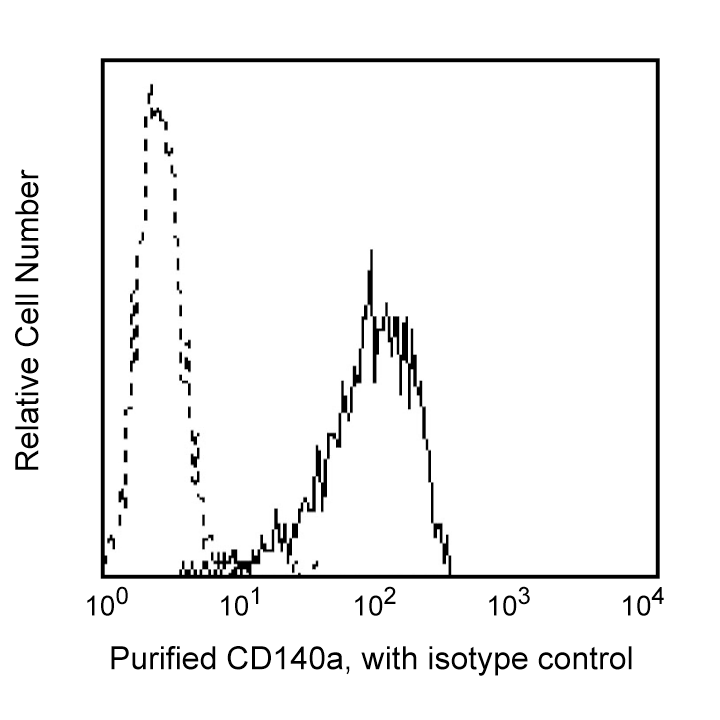
Flow cytometric analysis of MG-63 (osteosarcoma cell line) cells. MG-63 cells were stained with Purified Mouse Anti-Human CD140a (Cat. No. 556001; solid line histogram) or Purified Mouse IgG2a, κ Isotype Control (Cat. No. 555571; dashed line histogram), then FITC Goat Anti-Mouse IgG/IgM (Cat. No. 555988). The fluorescence histograms were derived from gated events with the forward and side light-scattering characteristics of viable MG-63 cells.

Flow cytometric analysis of MG-63 (osteosarcoma cell line) cells. MG-63 cells were stained with Purified Mouse Anti-Human CD140a (Cat. No. 556001; solid line histogram) or Purified Mouse IgG2a, κ Isotype Control (Cat. No. 555571; dashed line histogram), then FITC Goat Anti-Mouse IgG/IgM (Cat. No. 555988). The fluorescence histograms were derived from gated events with the forward and side light-scattering characteristics of viable MG-63 cells.


Preparation And Storage


Product Notices
- Since applications vary, each investigator should titrate the reagent to obtain optimal results.
- An isotype control should be used at the same concentration as the antibody of interest.
- Caution: Sodium azide yields highly toxic hydrazoic acid under acidic conditions. Dilute azide compounds in running water before discarding to avoid accumulation of potentially explosive deposits in plumbing.
- Sodium azide is a reversible inhibitor of oxidative metabolism; therefore, antibody preparations containing this preservative agent must not be used in cell cultures nor injected into animals. Sodium azide may be removed by washing stained cells or plate-bound antibody or dialyzing soluble antibody in sodium azide-free buffer. Since endotoxin may also affect the results of functional studies, we recommend the NA/LE (No Azide/Low Endotoxin) antibody format, if available, for in vitro and in vivo use.
- Please refer to www.bdbiosciences.com/us/s/resources for technical protocols.
Companion Products


The αR1 monoclonal antibody specifically binds to the human platelet derived growth factor (PDGF) receptor α (PDGFRα), also known as CD140a. CD140a is a 170 kDa single transmembrane glycoprotein expressed on fibroblasts, smooth muscle cells, glial cells and chondrocytes. PDGF receptors α and β are single glycoproteins with intracellular tyrosine kinase domains. They are structurally similar to the M-CSF receptor and CD117 (c-kit). Their ligand, PDGF, is a mitogen for connective tissue cells and glial cells. PDGF plays a role in wound healing and it also acts as a chemoattractant for fibroblasts, smooth muscle cells, glial cells, monocytes and neutrophils. Functional PDGF is secreted in disulfide linked, homodimeric or heterodimeric forms comprised of A or B chains (PDGFAA, PDGF-BB or PDGF-AB). Binding of divalent PDGF induces receptor dimerization with three possible forms: αα, αβ, ββ. The PDGFRα subunit binds both PDGF A and B chains, whereas the PDGFRβ subunit binds only PDGF B chains. Although both receptor subunits can stimulate mitogenic responses, only the β subunit can induce chemotaxis. The αR1 antibody is specific for PDGFRα and does not crossreact with PDGFRβ. It immunoprecipitates human, monkey, rabbit, pig, dog and cat PDGFRα. It does not recognize hamster, rat or mouse PDGFRα.
Development References (5)
-
Bazenet CE, Kazlauskas A. The PDGF receptor alpha subunit activates p21ras and triggers DNA synthesis without interacting with rasGAP. Oncogene. 1993; 9(2):517-525. (Biology). View Reference
-
Callard R, Gearing A. Callard R, Gearing A. The Cytokine Facts Book. San Diego: Academic Press; 1994.
-
Fitzgerald KA, Callard RE. The cytokine factsbook., 2nd ed. / Katherine A. Fitzgerald ... [et al.].. San Diego: Academic Press; 2001:1-515.
-
Kuroki M, Matsuo Y, Matsuoka Y. CD66 family Workshop: Reactivity of the CD66 Panel of monoclonal antibodies with soluble and membrane bound recombinant CD66 antigens. In: Kishimoto T. Tadamitsu Kishimoto .. et al., ed. Leucocyte typing VI : white cell differentiation antigens : proceedings of the sixth international workshop and conference held in Kobe, Japan, 10-14 November 1996. New York: Garland Pub.; 1997:1009-1010.
-
LaRochelle WJ, Jensen RA, Heidaran MA, et al. Inhibition of platelet-derived growth factor autocrine growth stimulation by a monoclonal antibody to the human alpha platelet-derived growth factor receptor. Cell Growth Differ. 1993; 4(7):547-553. (Clone-specific: Flow cytometry, Functional assay, Immunoprecipitation). View Reference
Please refer to Support Documents for Quality Certificates
Global - Refer to manufacturer's instructions for use and related User Manuals and Technical data sheets before using this products as described
Comparisons, where applicable, are made against older BD Technology, manual methods or are general performance claims. Comparisons are not made against non-BD technologies, unless otherwise noted.
For Research Use Only. Not for use in diagnostic or therapeutic procedures.