
-
Reagents
- Flow Cytometry Reagents
-
Western Blotting and Molecular Reagents
- Immunoassay Reagents
-
Single-Cell Multiomics Reagents
- BD® OMICS-Guard Sample Preservation Buffer
- BD® AbSeq Assay
- BD® OMICS-One Immune Profiler Protein Panel
- BD® Single-Cell Multiplexing Kit
- BD Rhapsody™ ATAC-Seq Assays
- BD Rhapsody™ Whole Transcriptome Analysis (WTA) Amplification Kit
- BD Rhapsody™ TCR/BCR Next Multiomic Assays
- BD Rhapsody™ Targeted mRNA Kits
- BD Rhapsody™ Accessory Kits
-
Functional Assays
-
Microscopy and Imaging Reagents
-
Cell Preparation and Separation Reagents
-
- BD® OMICS-Guard Sample Preservation Buffer
- BD® AbSeq Assay
- BD® OMICS-One Immune Profiler Protein Panel
- BD® Single-Cell Multiplexing Kit
- BD Rhapsody™ ATAC-Seq Assays
- BD Rhapsody™ Whole Transcriptome Analysis (WTA) Amplification Kit
- BD Rhapsody™ TCR/BCR Next Multiomic Assays
- BD Rhapsody™ Targeted mRNA Kits
- BD Rhapsody™ Accessory Kits
- Netherlands (English)
-
Change country/language
Old Browser
This page has been recently translated and is available in French now.
Looks like you're visiting us from {countryName}.
Would you like to stay on the current country site or be switched to your country?


LEFT: Western blot analysis of Sox9 in human hepatocellular carcinoma cells. After electrophoresis (SDS-PAGE) of Hep G2 cell lysate (Cat. No. 611555) in a 2D Tris-Glycine polyacrylamide gel, the proteins were transferred to PVDF membranes and then probed with Purified Mouse Anti-Sox9 (Cat. No. 565492) at concentrations of 0.25, 0.125, and 0.06 µg/ml (lanes 1, 2, and 3, respectively). Specific staining was detected with HRP Goat Anti-Mouse Ig (Cat No 554002). Sox9 is identified as a band of 62 kDa. MIDDLE: Flow cytometric analysis of SOX9 expression in human hepatocellular carcinoma cells. Hep G2 cells (ATCC HB-8065) were fixed with BD Cytofix™ Fixation Buffer (Cat. No. 554655), and permeabilized with BD Phosflow™ Perm Buffer III (Cat. No. 558050). The cells were stained with either Purified Mouse IgG1 κ Isotype Control (Cat. No. 554121; dashed line histogram) or Purified Mouse Anti-Sox9 (solid line histogram). The fluorescence histograms were derived from gated events with the forward and side light-scatter characteristics of intact Hep G2 cells. Flow cytometric analysis was performed using a BD FACSCanto™ II Flow Cytometer System. RIGHT: Immunofluorescent staining of SOX9 in human hepatocellular carcinoma cells. Hep G2 cells (ATCC HB-8065) were fixed with BD Cytofix™ Fixation Buffer (Cat. No. 554655), permeabilized with BD Phosflow™ Perm Buffer III (Cat. No. 558050), and stained with Purified Mouse Anti-Human Sox9 (pseudo-colored green) at 2.5 µg/ml. The second-step reagent was Alexa Fluor® 488 Goat Anti-Mouse Ig (Life Technologies), and the nuclear counter-staining was with BD Pharmingen™ DAPI Solution (Cat. No. 564907, pseudo-colored blue). The images were captured on a BD Pathway™ 435 Cell Analyzer and merged using BD Attovision™ software. Permeabilization with BD Perm/Wash™ (Cat. No. 554723) is not recommended for use with this antibody.


Immunohistochemical staining of Sox9 in human seminal vesicle (top row) and rat testis (bottom row). Following antigen retrieval with BD Retrievagen A buffer (Cat. no. 550524), the tissue sections were stained with either Purified Mouse IgG1 κ Isotype Control (Cat. No. 554121; left column) or Purified Mouse Anti-Sox9 (right column). A three-step staining procedure that employs Biotin Goat Anti-Mouse Ig polyclonal secondary antibody (Cat. No. 550337), HRP Streptavidin (Cat. No. 550946) and DAB (Cat. No. 550880) and Hematoxylin counterstaining were used. Original magnification: 20X (top row) and 40X (bottom row).


BD Pharmingen™ Purified Mouse Anti-Sox 9

BD Pharmingen™ Purified Mouse Anti-Sox 9

BD Pharmingen™ Purified Mouse Anti-Sox 9

BD Pharmingen™ Purified Mouse Anti-Sox 9

Regulatory Status Legend
Any use of products other than the permitted use without the express written authorization of Becton, Dickinson and Company is strictly prohibited.
Preparation And Storage
Product Notices
- Since applications vary, each investigator should titrate the reagent to obtain optimal results.
- Please refer to www.bdbiosciences.com/us/s/resources for technical protocols.
- Sodium azide is a reversible inhibitor of oxidative metabolism; therefore, antibody preparations containing this preservative agent must not be used in cell cultures nor injected into animals. Sodium azide may be removed by washing stained cells or plate-bound antibody or dialyzing soluble antibody in sodium azide-free buffer. Since endotoxin may also affect the results of functional studies, we recommend the NA/LE (No Azide/Low Endotoxin) antibody format, if available, for in vitro and in vivo use.
- Caution: Sodium azide yields highly toxic hydrazoic acid under acidic conditions. Dilute azide compounds in running water before discarding to avoid accumulation of potentially explosive deposits in plumbing.
- Alexa Fluor® is a registered trademark of Molecular Probes, Inc., Eugene, OR.
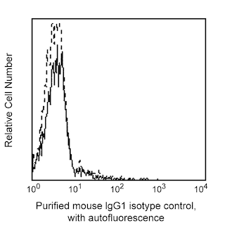
- An isotype control should be used at the same concentration as the antibody of interest.
Companion Products


The T32-668 monoclonal antibody specifically binds to Sox9, a member of the SRY-related HMG-box (SOX) family of transcription factors. Sox9 plays a key role in skeletal development and sex determination. Mutation of Sox9 has been associated with the human skeletal dysmorphology syndrome campomelic dysplasia, which is often characterized by both skeletal abnormalities and sex reversal. Expression of Sox9 has been observed in a variety of tissues and organs, including cartilage, testis, central neural system, lung, pancreas, intestinal stem cells, and heart.
Development References (5)
-
Furuyama K, Kawaguchi Y, Akiyama H, et al. Continuous cell supply from a Sox9-expressing progenitor zone in adult liver, exocrine pancreas and intestine. Nat Genet. 2011; 43(1):34-41. (Biology). View Reference
-
Guo W, Keckesova Z, Donaher JL, et al. Slug and Sox9 cooperatively determine the mammary stem cell state. Cell. 2012; 148(5):1015-1028. (Biology). View Reference
-
Kent J, Wheatley SC, Andrews JE, Sinclair AH, Koopman P. A male-specific role for SOX9 in vertebrate sex determination. Development. 1996; 122(9):2813-2822. (Biology). View Reference
-
Stolt CC, Lommes P, Sock E, Chaboissier MC, Schedl A, Wegner M. The Sox9 transcription factor determines glial fate choice in the developing spinal cord. Genes Dev. 2003; 17(13):1677-1689. (Biology). View Reference
-
Wright E, Hargrave MR, Christiansen J, et al. The Sry-related gene Sox9 is expressed during chondrogenesis in mouse embryos. Nat Genet. 1995; 9(1):15-20. (Biology). View Reference
Please refer to Support Documents for Quality Certificates
Global - Refer to manufacturer's instructions for use and related User Manuals and Technical data sheets before using this products as described
Comparisons, where applicable, are made against older BD Technology, manual methods or are general performance claims. Comparisons are not made against non-BD technologies, unless otherwise noted.
For Research Use Only. Not for use in diagnostic or therapeutic procedures.