Old Browser
This page has been recently translated and is available in French now.
Looks like you're visiting us from {countryName}.
Would you like to stay on the current country site or be switched to your country?


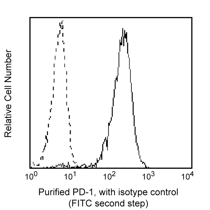
Profile of anti-PD-1 (MIH4) reactivity on MIH34 transfectant cells analyzed by flow cytometry. Second step staining with Cat. No. 555988.


BD Pharmingen™ Purified Mouse Anti-Human CD279

Regulatory Status Legend
Any use of products other than the permitted use without the express written authorization of Becton, Dickinson and Company is strictly prohibited.
Preparation And Storage
Product Notices
- Please refer to www.bdbiosciences.com/us/s/resources for technical protocols.
- Caution: Sodium azide yields highly toxic hydrazoic acid under acidic conditions. Dilute azide compounds in running water before discarding to avoid accumulation of potentially explosive deposits in plumbing.
- Since applications vary, each investigator should titrate the reagent to obtain optimal results.
Companion Products

.png?imwidth=320)
The MIH4 monoclonal antibody specifically binds to CD279, which is also known as, Programmed cell death 1 (PD-1). CD279 is a type I transmembrane glycoprotein that belongs to the Ig superfamily. CD279 is an immunoregulatory receptor that is expressed on expressed on subsets of thymocytes, activated T cells, B cells and myeloid cells. CD279 contains an immunoreceptor tyrosine-based inhibitory motif (ITIM) in its cytoplasmic region. CD273 (PD-L2) and CD274 (PD-L1) are ligands of CD279 and are members of the B7 gene family. Interaction of CD279 with its ligands results in inhibition of T cell proliferation and cytokine secretion. CD279 may play roles in supporting self-tolerance, reducing autoimmunity, or promoting T cell exhaustion associated with certain diseases.
Development References (5)
-
Bennett F, Luxenberg D, Ling V, et al. Program death-1 engagement upon TCR activation has distinct effects on costimulation and cytokine-driven proliferation: attenuation of ICOS, IL-4, and IL-21, but not CD28, IL-7, and IL-15 responses. J Immunol. 2003; 170(2):711-718. (Biology). View Reference
-
Carter L, Fouser LA, Jussif J, et al. PD-1:PD-L inhibitory pathway affects both CD4(+) and CD8(+) T cells and is overcome by IL-2. Eur J Immunol. 2002; 32:634-643. (Biology). View Reference
-
Freeman GJ, Long AJ, Iwai Y, et al. Engagement of PD-1 immunoinhibitory receptor by a novel B7 family member leads to negative regulation of lymphocyte activation. J Exp Med. 2000; 192:1027-1034. (Biology). View Reference
-
Latchman Y, Wood CR, Chernova T, et al. PD-L2 is a second ligand for PD-1 and inhibits T cell activation. Nat Immunol. 2001; 2(3):261-268. (Biology). View Reference
-
Nishimura H, Minato N, Nakano T, Honjo T. Immunological studies on PD-1 deficient mice: implication of PD-1 as a negative regulator for B cell responses. Int Immunol. 1998; 10(10):1563-1572. (Biology). View Reference
Please refer to Support Documents for Quality Certificates
Global - Refer to manufacturer's instructions for use and related User Manuals and Technical data sheets before using this products as described
Comparisons, where applicable, are made against older BD Technology, manual methods or are general performance claims. Comparisons are not made against non-BD technologies, unless otherwise noted.
For Research Use Only. Not for use in diagnostic or therapeutic procedures.
Report a Site Issue
This form is intended to help us improve our website experience. For other support, please visit our Contact Us page.