





.png)


Preparation And Storage


Product Notices
- This reagent has been pre-diluted for use at the recommended Volume per Test. We typically use 1 × 10^6 cells in a 100-µl experimental sample (a test).
- An isotype control should be used at the same concentration as the antibody of interest.
- Source of all serum proteins is from USDA inspected abattoirs located in the United States.
- Caution: Sodium azide yields highly toxic hydrazoic acid under acidic conditions. Dilute azide compounds in running water before discarding to avoid accumulation of potentially explosive deposits in plumbing.
- Please observe the following precautions: Absorption of visible light can significantly alter the energy transfer occurring in any tandem fluorochrome conjugate; therefore, we recommend that special precautions be taken (such as wrapping vials, tubes, or racks in aluminum foil) to prevent exposure of conjugated reagents, including cells stained with those reagents, to room illumination.
- For fluorochrome spectra and suitable instrument settings, please refer to our Multicolor Flow Cytometry web page at www.bdbiosciences.com/colors.
- Texas Red is a registered trademark of Molecular Probes, Inc., Eugene, OR.
- CF™ is a trademark of Biotium, Inc.
- When excited by the yellow-green (561-nm) laser, the fluorescence may be brighter than when excited by the blue (488-nm) laser.
- This product is provided under an Agreement between BIOTIUM and BD Biosciences. The manufacture, use, sale, offer for sale, or import of this product is subject to one or more patents or pending applications owned or licensed by Biotium, Inc. This product, and only in the amount purchased by buyer, may be used solely for buyer’s own internal research, in a manner consistent with the accompanying product literature. No other right to use, sell or otherwise transfer (a) this product, or (b) its components is hereby granted expressly, by implication or by estoppel. This product is for research use only. Diagnostic uses require a separate license from Biotium, Inc. For information on purchasing a license to this product including for purposes other than research, contact Biotium, Inc., 3159 Corporate Place, Hayward, CA 94545, Tel: (510) 265-1027. Fax: (510) 265-1352. Email: btinfo@biotium.com.
- Because of the broad absorption spectrum of the tandem fluorochrome, extra care must be taken when using multi-laser cytometers, which may directly excite both PE and CF™594.
- Please refer to http://regdocs.bd.com to access safety data sheets (SDS).
- Species cross-reactivity detected in product development may not have been confirmed on every format and/or application.
- Please refer to www.bdbiosciences.com/us/s/resources for technical protocols.
Companion Products
.png?imwidth=320)
The JES3-19F1 monoclonal antibody specifically recognizes human Interleukin-10 (IL-10) that is encoded by IL10. IL-10 is also known as Cytokine Synthesis Inhibitory Factor (CSIF), B cell-derived T cell growth factor (B-TCGF), and T-cell growth inhibitory factor (TGIF). The JES3-19F1 antibody crossreacts with ebvIL-10 protein, the Epstein-Barr viral IL-10 homolog (viral IL-10 or vIL-10) encoded by the BCRF1 gene. IL-10 is produced by a variety of cells such as some activated T cells and B cells including regulatory T cells (Treg) and B cells (Breg), monocytes and macrophages, dendritic cells (DC), keratinocytes, and mast cells. IL-10 is a multifunctional cytokine that can downregulate immune and proinflammatory responses. IL-10 can act to reduce expression of major histocompatibility complex class II antigens, costimulatory molecules, or proinflammatory cytokines including IL-1β, IL-2, IL-3, IL-12, IFN-γ, TNF or GM-CSF expressed by activated monocytes, macrophages, dendritic cells (DC), natural killer (NK) cells, or T cells. IL-10 has been shown to play a role in chronic viral infections. IL-10 can also enhance B cell survival, proliferation, and differentiation to become antibody-producing cells. The JES3-19F1 antibody reportedly neutralizes the biological activity of human IL-10 and ebvIL-10. IL-10 mediates its biological activities by signaling through a heterotetrameric receptor complex composed of the type II cytokine receptor subunits CD210a (IL-10 Rα) and CD210b (IL-10 Rβ).
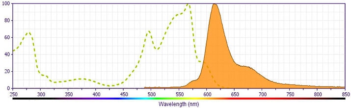
This antibody is conjugated to BD Horizon PE-CF594, which has been developed exclusively by BD Biosciences as a better alternative to PE-Texas Red®. PE-CF594 excites and emits at similar wavelengths to PE-Texas Red® yet exhibits improved brightness and spectral characteristics. Due to PE having maximal absorption peaks at 496 nm and 564 nm, PE-CF594 can be excited by the blue (488-nm), green (532-nm) and yellow-green (561-nm) lasers and can be detected with the same filter set as PE-Texas Red® (eg, 610/20-nm filter).
Development References (5)
-
Abrams JS, Roncarolo MG, Yssel H, Andersson U, Gleich GJ, Silver JE. Strategies of anti-cytokine monoclonal antibody development: immunoassay of IL-10 and IL-5 in clinical samples. Immunol Rev. 1992; 127:5-24. (Clone-specific: ELISA, ELISPOT, Flow cytometry, Neutralization). View Reference
-
Bacchetta R, Bigler M, Touraine JL, et al. High levels of interleukin 10 production in vivo are associated with tolerance in SCID patients transplanted with HLA mismatched hematopoietic stem cells. J Exp Med. 1994; 179(2):493-502. (Clone-specific: Neutralization). View Reference
-
D'Andrea A, Aste-Amezaga M, Valiante NM, Ma X, Kubin M, Trinchieri G. Interleukin 10 (IL-10) inhibits human lymphocyte interferon gamma-production by suppressing natural killer cell stimulatory factor/IL-12 synthesis in accessory cells. J Exp Med. 1993; 178(3):1041-1048. (Clone-specific: Neutralization). View Reference
-
Prussin C, Metcalfe DD. Detection of intracytoplasmic cytokine using flow cytometry and directly conjugated anti-cytokine antibodies. J Immunol Methods. 1995; 188(1):117-128. (Methodology: Flow cytometry). View Reference
-
Schlaak JF, Schmitt E, Hüls C, Meyer zum Büschenfelde KH, Fleischer B. A sensitive and specific bioassay for the detection of human interleukin-10. J Immunol Methods. 1994; 168(1):49-54. (Biology). View Reference
Please refer to Support Documents for Quality Certificates
Global - Refer to manufacturer's instructions for use and related User Manuals and Technical data sheets before using this products as described
Comparisons, where applicable, are made against older BD Technology, manual methods or are general performance claims. Comparisons are not made against non-BD technologies, unless otherwise noted.
For Research Use Only. Not for use in diagnostic or therapeutic procedures.
