Old Browser
This page has been recently translated and is available in French now.
Looks like you're visiting us from {countryName}.
Would you like to stay on the current country site or be switched to your country?


Flow cytometric analysis of CCRL1 expression on human Hep 3B2 cells. Cells from the human Hep 3B2 (Hepatocellular carcinoma, ATCC HB-8064) cell line were stained with either Mouse IgG2b κ Isotype Control (Cat. No. 562748; dotted line histogram) or BD Horizon™ BV421 Mouse Anti-Human CCRL1 antibody (Cat. No. 566601; solid line histogram) at 0.5 µg/test. The fluorescence histogram showing CCRL1 expression (or Ig Isotype control staining) was derived from gated events with the forward and side light-scatter characteristics of viable cells. Flow cytometric analysis was performed using a BD FACSCanto™ II Flow Cytometer System. Data shown on this Technical Data Sheet are not lot specific.

Immunohistofluorescent staining of CCRL1 on human cardiac muscle cells. A frozen human cardiac muscle section was fixed with cold acetone, washed, and then stained with BV421 Mouse Anti-Human CCRL1 antibody (Cat. No. 566601). The cells showed positive membrane staining for CCRL1 (pseudocolored red) and counterstaining (pseudocolored green) with DRAQ5™ (Cat. No. 564902/564903). The image was acquired using a Molecular Devices Epifluorescence microscope. Original magnification, 20×.


BD Horizon™ BV421 Mouse Anti-Human CCRL1

BD Horizon™ BV421 Mouse Anti-Human CCRL1

Regulatory Status Legend
Any use of products other than the permitted use without the express written authorization of Becton, Dickinson and Company is strictly prohibited.
Preparation And Storage
Recommended Assay Procedures
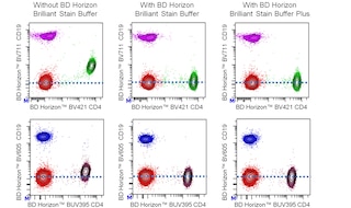
For optimal and reproducible results, BD Horizon Brilliant Stain Buffer should be used anytime two or more BD Horizon Brilliant dyes are used in the same experiment. Fluorescent dye interactions may cause staining artifacts which may affect data interpretation. The BD Horizon Brilliant Stain Buffer was designed to minimize these interactions. More information can be found in the Technical Data Sheet for the BD Horizon Brilliant Stain Buffer (Cat. No. 563794/566349) or the BD Horizon Brilliant Stain Buffer Plus (Cat. No. 566385).
Product Notices
- Please refer to www.bdbiosciences.com/us/s/resources for technical protocols.
- Source of all serum proteins is from USDA inspected abattoirs located in the United States.
- Caution: Sodium azide yields highly toxic hydrazoic acid under acidic conditions. Dilute azide compounds in running water before discarding to avoid accumulation of potentially explosive deposits in plumbing.
- For fluorochrome spectra and suitable instrument settings, please refer to our Multicolor Flow Cytometry web page at www.bdbiosciences.com/colors.
- Pacific Blue™ is a trademark of Molecular Probes, Inc., Eugene, OR.
- BD Horizon Brilliant Violet 421 is covered by one or more of the following US patents: 8,158,444; 8,362,193; 8,575,303; 8,354,239.
- BD Horizon Brilliant Stain Buffer is covered by one or more of the following US patents: 8,110,673; 8,158,444; 8,575,303; 8,354,239.
- An isotype control should be used at the same concentration as the antibody of interest.
- Since applications vary, each investigator should titrate the reagent to obtain optimal results.
Companion Products


The 13E11 monoclonal antibody specifically binds to CC chemokine receptor-like 1 (CCRL1) that is also known as C-C chemokine receptor type 11 (C-C CKR-11), or ChemoCentryx chemokine receptor (CCX-CKR). CCRL1 is a seven-transmembrane glycoprotein receptor that is encoded by ACKR4 (Atypical chemokine receptor 4) which belongs to the atypical chemokine receptor subfamily within the chemokine receptor superfamily. CCRL1 is expressed on stromal cells of lymph nodes, dermal lymphatic endothelial cells, and thymic epithelial cells. CCRL1 binds to several "homeostatic" chemokines including CCL19 (ELC), CCL21 (SLC), CCL25 (TECK) and CXCL13 (BLC) that can otherwise bind to functional chemokine receptors like CCR7, CCR9 or CXCR5. CCRL1 can thus serve as a decoy receptor that allows cellular uptake and degradation of these chemokines in a G protein-independent manner. This receptor may shape chemokine gradients in tissues by scavenging chemokines and thereby regulate the chemotaxis of thymic precursor cells, various leucocytes, including lymphocytes and dendritic cells, and cancer cells. CCRL1 expression is upregulated on some breast and hepatic cancer cell lines. The 13E11 antibody reportedly has ligand-like activity that results in the cellular internalization of CCRL1. This antibody recognizes an epitope in the N-terminus of the first extracellular domain of CCRL1.
The antibody was conjugated to BD Horizon BV421 which is part of the BD Horizon Brilliant™ Violet family of dyes. With an Ex Max of 407-nm and Em Max at 421-nm, BD Horizon BV421 can be excited by the violet laser and detected in the standard Pacific Blue™ filter set (eg, 450/50-nm filter). BD Horizon BV421 conjugates are very bright, often exhibiting a 10 fold improvement in brightness compared to Pacific Blue conjugates.

Development References (4)
-
Bachelerie F, Ben-Baruch A, Burkhardt AM, et al. International Union of Basic and Clinical Pharmacology. [corrected]. LXXXIX. Update on the extended family of chemokine receptors and introducing a new nomenclature for atypical chemokine receptors.. Pharmacol Rev. 2014; 66(1):1-79. (Biology). View Reference
-
Bryce SA, Wilson RA, Tiplady EM, et al. ACKR4 on Stromal Cells Scavenges CCL19 To Enable CCR7-Dependent Trafficking of APCs from Inflamed Skin to Lymph Nodes.. J Immunol. 2016; 196(8):3341-53. (Clone-specific: Fluorescence microscopy, Immunofluorescence). View Reference
-
Takatsuka S, Sekiguchi A, Tokunaga M, Fujimoto A, Chiba J. Generation of a panel of monoclonal antibodies against atypical chemokine receptor CCX-CKR by DNA immunization.. J Pharmacol Toxicol Methods. 63(3):250-7. (Immunogen: Blocking, Cytotoxicity, Flow cytometry, Fluorescence microscopy, Functional assay, Immunofluorescence). View Reference
-
Watts AO, Verkaar F, van der Lee MM, et al. β-Arrestin recruitment and G protein signaling by the atypical human chemokine decoy receptor CCX-CKR.. J Biol Chem. 2013; 288(10):7169-81. (Biology). View Reference
Please refer to Support Documents for Quality Certificates
Global - Refer to manufacturer's instructions for use and related User Manuals and Technical data sheets before using this products as described
Comparisons, where applicable, are made against older BD Technology, manual methods or are general performance claims. Comparisons are not made against non-BD technologies, unless otherwise noted.
For Research Use Only. Not for use in diagnostic or therapeutic procedures.
Report a Site Issue
This form is intended to help us improve our website experience. For other support, please visit our Contact Us page.