
Preparation And Storage
Product Notices
- This reagent has been pre-diluted for use at the recommended Volume per Test. We typically use 1 × 10^6 cells in a 100-µl experimental sample (a test).
- An isotype control should be used at the same concentration as the antibody of interest.
- The Alexa Fluor®, Pacific Blue™, and Cascade Blue® dye antibody conjugates in this product are sold under license from Molecular Probes, Inc. for research use only, excluding use in combination with microarrays, or as analyte specific reagents. The Alexa Fluor® dyes (except for Alexa Fluor® 430), Pacific Blue™ dye, and Cascade Blue® dye are covered by pending and issued patents.
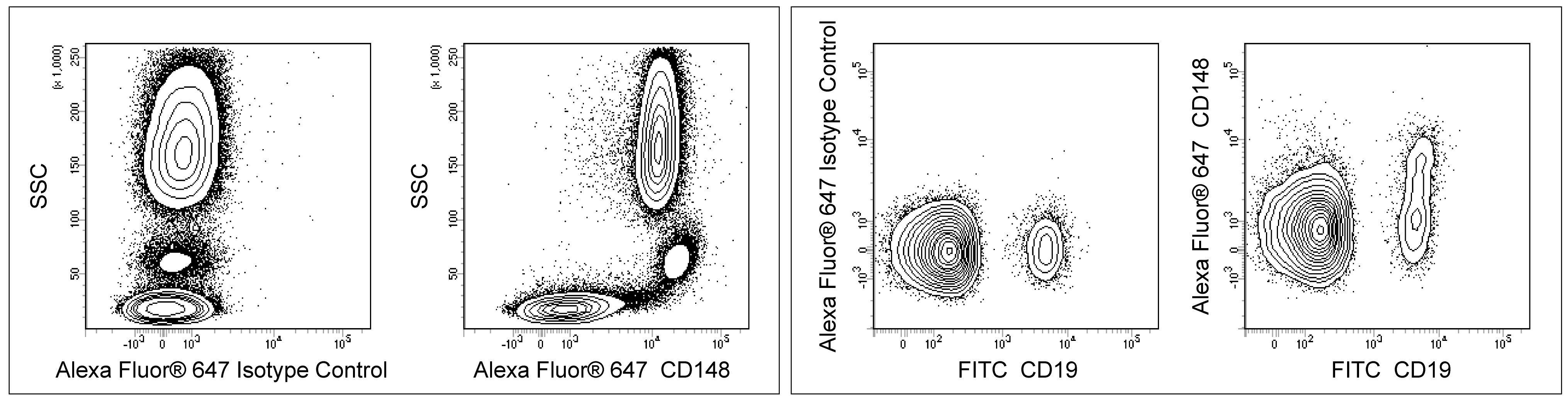
- Alexa Fluor® 647 fluorochrome emission is collected at the same instrument settings as for allophycocyanin (APC).
- Alexa Fluor® is a registered trademark of Molecular Probes, Inc., Eugene, OR.
- Caution: Sodium azide yields highly toxic hydrazoic acid under acidic conditions. Dilute azide compounds in running water before discarding to avoid accumulation of potentially explosive deposits in plumbing.
- For fluorochrome spectra and suitable instrument settings, please refer to our Multicolor Flow Cytometry web page at www.bdbiosciences.com/colors.
- Source of all serum proteins is from USDA inspected abattoirs located in the United States.
- Please refer to www.bdbiosciences.com/us/s/resources for technical protocols.
Companion Products


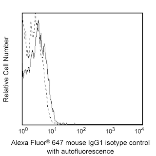
The A3 monoclonal antibody specifically binds to CD148. CD148 is a receptor-type tyrosine phosphatase that belongs to the receptor class 3 subfamily of the protein-tyrosine phosphatase family. CD148 is also known as Density-enhanced phosphatase 1 (DEP-1), Human protein tyrosine phosphatase- η (HPTP-η/HPTP-eta), or Protein tyrosine phosphatase receptor type J (PTPRJ). CD148 is a highly glycosylated type I transmembrane protein that is widely expressed on different cell types including monocytes, granulocytes, dendritic cells, thymocytes, T cells, B cells, NK cells, fibroblasts, and platelets. As a protein tyrosine phosphatase with receptor function, CD148 plays a role in regulating the signaling activities of various phosphorylated cellular proteins including the PDGF Receptor, catenins and Src family kinases. In the immune system, CD148 plays a role in regulating the activities of different cell types. For example, upon T cell activation, CD148 expression is upregulated by T cells. CD148 can subsequently dephosphorylate LAT and PLC-γ signaling proteins and thereby downregulate T cell signaling responses. Immobilized A3 antibody can reportedly augment the proliferation of anti-CD3 antibody-stimulated peripheral blood T cells in culture.
Development References (4)
-
Baker JE, Majeti R, Tangye SG, Weiss A. Protein tyrosine phosphatase CD148-mediated inhibition of T-cell receptor signal transduction is associated with reduced LAT and phospholipase Cgamma1 phosphorylation. Mol Cell Biol. 2001; 21(7):2393-2403. (Clone-specific: Flow cytometry). View Reference
-
Scraven B, Hegen M, Autschbach F, Gaya A, Schwarz C, Meuer SC. CD148 (p260 phosphatase) Workshop Panel report. In: Kishimoto T. Tadamitsu Kishimoto .. et al., ed. Leucocyte typing VI : white cell differentiation antigens : proceedings of the sixth international workshop and conference held in Kobe, Japan, 10-14 November 1996. New York: Garland Pub.; 1997:576-580.
-
Tangye SG, Phillips JH, Lanier LL, de Vries JE, Aversa G. CD148: a receptor-type protein tyrosine phosphatase involved in the regulation of human T cell activation. J Immunol. 1998; 161(7):9249-9255. (Immunogen: (Co)-stimulation, Flow cytometry, Functional assay). View Reference
-
Whiteford JR, Xian X, Chaussade C, Vanhaesebroeck B, Nourshargh S, Couchman JR. Syndecan-2 is a novel ligand for the protein tyrosine phosphatase receptor CD148. Mol Cell Biol. 2011; 22(19):3609-3624. (Clone-specific: Flow cytometry, Functional assay, Western blot). View Reference
Please refer to Support Documents for Quality Certificates
Global - Refer to manufacturer's instructions for use and related User Manuals and Technical data sheets before using this products as described
Comparisons, where applicable, are made against older BD Technology, manual methods or are general performance claims. Comparisons are not made against non-BD technologies, unless otherwise noted.
For Research Use Only. Not for use in diagnostic or therapeutic procedures.