







Preparation And Storage


Product Notices
- Since applications vary, each investigator should titrate the reagent to obtain optimal results.
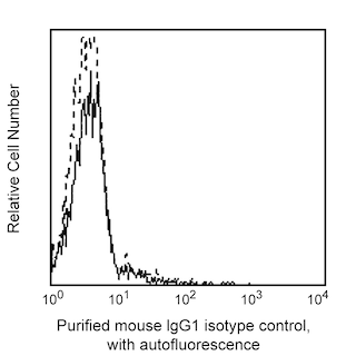
- An isotype control should be used at the same concentration as the antibody of interest.
- Caution: Sodium azide yields highly toxic hydrazoic acid under acidic conditions. Dilute azide compounds in running water before discarding to avoid accumulation of potentially explosive deposits in plumbing.
- Please refer to http://regdocs.bd.com to access safety data sheets (SDS).
- Species cross-reactivity detected in product development may not have been confirmed on every format and/or application.
- Sodium azide is a reversible inhibitor of oxidative metabolism; therefore, antibody preparations containing this preservative agent must not be used in cell cultures nor injected into animals. Sodium azide may be removed by washing stained cells or plate-bound antibody or dialyzing soluble antibody in sodium azide-free buffer. Since endotoxin may also affect the results of functional studies, we recommend the NA/LE (No Azide/Low Endotoxin) antibody format, if available, for in vitro and in vivo use.
- Please refer to www.bdbiosciences.com/us/s/resources for technical protocols.
Companion Products
.png?imwidth=320)
The HIP8 monoclonal antibody specifically binds to the α-chain of CD41. CD41 is also known as Integrin αIIb or Platelet GPIIb. The calcium-dependent complex of CD41 and CD61 (β3 integrin or GPIIIa) is normally expressed on platelets and megakaryocytes. The CD41/CD61 complex is the receptor for fibrinogen, fibronectin and von Willebrand factor, and mediates platelet adhesion and aggregation. CD41 (clone HIP8) completely inhibits ADP-, epinephrine-, and collagen-induced platelet activation, and partially inhibits ristocetin- and thrombin-induced platelet activation. This antibody is useful in the morphological and physiological studies of platelets and megakaryocytes.
Clone HIP8 also cross-reacts with a major subset of peripheral blood platelets of baboon, and both rhesus and cynomolgus macaque monkeys. The staining pattern of platelets is similar to that observed with peripheral blood platelets from normal human donors.
Development References (2)
-
Knapp W. W. Knapp .. et al., ed. Leucocyte typing IV : white cell differentiation antigens. Oxford New York: Oxford University Press; 1989:1-1182.
-
Schlossman SF. Stuart F. Schlossman .. et al., ed. Leucocyte typing V : white cell differentiation antigens : proceedings of the fifth international workshop and conference held in Boston, USA, 3-7 November, 1993. Oxford: Oxford University Press; 1995.
Please refer to Support Documents for Quality Certificates
Global - Refer to manufacturer's instructions for use and related User Manuals and Technical data sheets before using this products as described
Comparisons, where applicable, are made against older BD Technology, manual methods or are general performance claims. Comparisons are not made against non-BD technologies, unless otherwise noted.
For Research Use Only. Not for use in diagnostic or therapeutic procedures.
