
Preparation And Storage
Recommended Assay Procedures
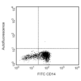
BD® CompBeads can be used as surrogates to assess fluorescence spillover (compensation). When fluorochrome conjugated antibodies are bound to BD® CompBeads, they have spectral properties very similar to cells. However, for some fluorochromes there can be small differences in spectral emissions compared to cells, resulting in spillover values that differ when compared to biological controls. It is strongly recommended that when using a reagent for the first time, users compare the spillover on cell and BD® CompBeads to ensure that BD® CompBeads are appropriate for your specific cellular application.
Product Notices
- Please refer to www.bdbiosciences.com/us/s/resources for technical protocols.
- This reagent has been pre-diluted for use at the recommended Volume per Test. We typically use 1 × 10^6 cells in a 100-µl experimental sample (a test).
- An isotype control should be used at the same concentration as the antibody of interest.
- Source of all serum proteins is from USDA inspected abattoirs located in the United States.
- Caution: Sodium azide yields highly toxic hydrazoic acid under acidic conditions. Dilute azide compounds in running water before discarding to avoid accumulation of potentially explosive deposits in plumbing.
- For fluorochrome spectra and suitable instrument settings, please refer to our Multicolor Flow Cytometry web page at www.bdbiosciences.com/colors.
- Please refer to http://regdocs.bd.com to access safety data sheets (SDS).
Companion Products


The 4H9 (Leu-9) monoclonal antibody specifically recognizes human CD7 which is also known as T-cell leukemia antigen, T-cell surface antigen Leu-9, and LEU-9. CD7 is a ~40 kDa type I transmembrane glycoprotein that has an extracellular region with an N-terminal IgV-like domain followed by an extended O-glycosylated stalk region, a transmembrane region and cytoplasmic tail. The CD7 antigen is expressed throughout T-lymphocyte differentiation. It is present on 85% to 90% of peripheral blood T lymphocytes. In normal individuals, CD7 is expressed on all CD8+ lymphocytes, approximately 90% of CD4+ lymphocytes, and most NK cells. CD7 is weakly expressed on monocytes but not on granulocytes or B lymphocytes. It is expressed on hematopoietic progenitors and 50% of thymocytes. In leukemias, the CD7 antigen is present on the majority of T-lymphoid lineages. CD7 may function in cellular adhesion and play a role in interactions between T cells as well as T cells and B cells.

Development References (9)
-
Foon KA, Todd RF. Immunologic classification of leukemia and lymphoma.. Blood. 1986; 68(1):1-31. (Clone-specific). View Reference
-
Link M, Warnke R, Finlay J, et al. A single monoclonal antibody identifies T-cell lineage of childhood lymphoid malignancies.. Blood. 1983; 62(4):722-8. (Immunogen: Flow cytometry). View Reference
-
Palker TJ, Scearce RM, Hensley LL, Ho W, Haynes BF. Comparison of the CD7 (3A1) group of T cell workshop antibodies. In: Reinherz EL, Haynes BF, Nadler LM, Bernstein ID, ed. Leukocyte Typing II. Human T Lymphocytes. New York, NY: Springer-Verlag; 1986:303-313.
-
Picker LJ, Weiss LM, Medeiros LJ, Wood GS, Warnke RA. Immunophenotypic criteria for the diagnosis of non-Hodgkin's lymphoma.. Am J Pathol. 1987; 128(1):181-201. (Clone-specific: Immunohistochemistry). View Reference
-
Rabinowich H, Pricop L, Herberman RB, Whiteside TL. Expression and function of CD7 molecule on human natural killer cells. J Immunol. 1994; 152(2):517-526. (Clone-specific: Flow cytometry). View Reference
-
Weiss LM, Crabtree GS, Rouse RV, Warnke RA. Morphologic and immunologic characterization of 50 peripheral T-cell lymphomas.. Am J Pathol. 1985; 118(2):316-24. (Clone-specific: Immunohistochemistry). View Reference
-
Weiss LM, Wood GS, Warnke RA. Immunophenotypic differences between dermatopathic lymphadenopathy and lymph node involvement in mycosis fungoides.. Am J Pathol. 1985; 120(2):179-85. (Clone-specific: Immunohistochemistry). View Reference
-
Wood GS, Abel EA, Hoppe RT, Warnke RA. Leu-8 and Leu-9 antigen phenotypes: immunologic criteria for the distinction of mycosis fungoides from cutaneous inflammation.. J Am Acad Dermatol. 1986; 14(6):1006-13. (Clone-specific: Immunohistochemistry). View Reference
-
Zola H, Swart B, Nicholson I, Voss E. CD7. In: Zola H. Leukocyte and stromal cell molecules : the CD markers. Hoboken, N.J.: Wiley-Liss; 2007:52.
Please refer to Support Documents for Quality Certificates
Global - Refer to manufacturer's instructions for use and related User Manuals and Technical data sheets before using this products as described
Comparisons, where applicable, are made against older BD Technology, manual methods or are general performance claims. Comparisons are not made against non-BD technologies, unless otherwise noted.
For Research Use Only. Not for use in diagnostic or therapeutic procedures.