
Preparation And Storage
Recommended Assay Procedures
Flow Cytometric Analysis: The PE-Cy™7 Mouse Anti-Human IFN-γ antibody is useful for immunofluorescent staining and flow cytometric analysis to identify and enumerate IFN-γ producing cells within mixed cell populations. For specific methodology, please visit our web site, http://www.bdbiosciences.com/us/s/resources, and go to the protocols section under "Intracellular Staining".
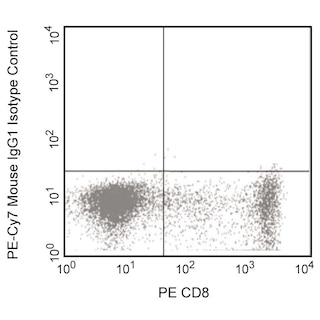
A useful control for demonstrating specificity of staining is pre-block the fixed/permeabilized cells with Purified Mouse Anti-Human IFN-γ (Cat. No. 554549) prior to staining. The staining technique and blocking controls are described in detail by C. Prussin and D. Metcalfe. A suitable mouse IgG1 isotype control for assessing the level of background staining on paraformaldehyde-fixed/saponin-permeabilized human cells is PE-Cy™7 Mouse IgG1¦Isotype Control (Cat. No. 557646); use at comparable concentrations to antibody of interest (e.g., ¡ 0.5 ¦g mAb/1 million cells).
Product Notices
- This reagent has been pre-diluted for use at the recommended Volume per Test. We typically use 1 × 10^6 cells in a 100-µl experimental sample (a test).
- An isotype control should be used at the same concentration as the antibody of interest.
- Source of all serum proteins is from USDA inspected abattoirs located in the United States.
- Caution: Sodium azide yields highly toxic hydrazoic acid under acidic conditions. Dilute azide compounds in running water before discarding to avoid accumulation of potentially explosive deposits in plumbing.
- Warning: Some APC-Cy7 and PE-Cy7 conjugates show changes in their emission spectrum with prolonged exposure to formaldehyde. If you are unable to analyze fixed samples within four hours, we recommend that you use BD™ Stabilizing Fixative (Cat. No. 338036).
- Please observe the following precautions: Absorption of visible light can significantly alter the energy transfer occurring in any tandem fluorochrome conjugate; therefore, we recommend that special precautions be taken (such as wrapping vials, tubes, or racks in aluminum foil) to prevent exposure of conjugated reagents, including cells stained with those reagents, to room illumination.
- PE-Cy7 is a tandem fluorochrome composed of R-phycoerythrin (PE), which is excited by 488-nm light and serves as an energy donor, coupled to the cyanine dye Cy7, which acts as an energy acceptor and fluoresces maximally at 780 nm. PE-Cy7 tandem fluorochrome emission is collected in a detector for fluorescence wavelengths of 750 nm and higher. Although every effort is made to minimize the lot-to-lot variation in the efficiency of the fluorochrome energy transfer, differences in the residual emission from PE may be observed. Therefore, we recommend that individual compensation controls be performed for every PE-Cy7 conjugate. PE-Cy7 is optimized for use with a single argon ion laser emitting 488-nm light, and there is no significant overlap between PE-Cy7 and FITC emission spectra. When using dual-laser cytometers, which may directly excite both PE and Cy7, we recommend the use of cross-beam compensation during data acquisition or software compensation during data analysis.
- For fluorochrome spectra and suitable instrument settings, please refer to our Multicolor Flow Cytometry web page at www.bdbiosciences.com/colors.
- Species cross-reactivity detected in product development may not have been confirmed on every format and/or application.
- Cy is a trademark of GE Healthcare.
- Please refer to http://regdocs.bd.com to access safety data sheets (SDS).
- Please refer to www.bdbiosciences.com/us/s/resources for technical protocols.
Companion Products


The 4S.B3 monoclonal antibody specifically binds to interferon-γ (IFN-γ). The immunogen used to generate this hybridoma was partially purified human IFN-γ obtained from supernatants of human PBMC stimulated with Staphylococcus aureus. Interferon-γ (IFN-γ) is a potent multifunctional cytokine that is produced by several activated cell types including NK, NKT, CD4+TCRαβ+, CD8+TCRαβ+, and TCRγδ+ T cells. IFN-γ exerts its biological effects through specific binding to the high-affinity IFN-γ Receptor Complex comprised of IFN-γRα (CD119) and IFN-γRβ subunits. In addition to its antiviral effects, IFN-γ upregulates a number of lymphoid cell functions including the antimicrobial and antitumor responses of macrophages, NK cells, and neutrophils. In addition, IFN-γ can exert strong regulatory influences on the proliferation, differentiation, and effector responses of B cell and T cell subsets. These influences can involve IFN-γ's capacity to boost MHC class I and II expression by antigen-presenting cells as well as to direct effects on B cells and T cells themselves. Human IFN-γ is a 14-18 kDa glycoprotein containing 143 amino acid residues.
Clone 4S.B3 also cross-reacts with a cytoplasmic component of peripheral blood CD3+ lymphocytes of baboon, and both rhesus and cynomolgus macaque monkeys following five-hour treatment with phorbol myristic acetate (PMA) and Ca++ ionophore (A23187) in the presence of monensin. The staining pattern of 4S.B3 in CD3+ cells is similar to that observed with peripheral blood T lymphocytes from normal human donors. This reagent is useful for intracellular immunofluorescent staining for flow cytometric analysis to identify and enumerate IFN-γ + cells within a mixed cell population.

Development References (6)
-
Farrar MA, Schreiber RD. The molecular cell biology of interferon-gamma and its receptor. Annu Rev Immunol. 1993; 11:571-611. (Biology). View Reference
-
Favre C, Wijdenes J, Cabrillat H, Djossou O, Banchereau J, de Vries JE. Epitope mapping of recombinant human gamma interferon using monoclonal antibodies. Mol Immunol. 1989; 26(1):17-25. (Biology). View Reference
-
Meager A. Characterization of interferons and immunoassays. In: Clemens MJ, Morris AG, Gearing AJH, ed. Lymphokines and Interferons. A Practical Approach. Oxford: IRL Press Ltd; 1987:105-127.
-
Prussin C, Metcalfe DD. Detection of intracytoplasmic cytokine using flow cytometry and directly conjugated anti-cytokine antibodies. J Immunol Methods. 1995; 188(1):117-128. (Methodology: Flow cytometry). View Reference
-
Sester U, Sester M, Hauk M, Kaul H, Köhler H, Girndt M. T-cell activation follows Th1 rather than Th2 pattern in haemodialysis patients.. Nephrol Dial Transplant. 2000; 15(8):1217-1223. (Biology). View Reference
-
Vogel S, Friedman R, Hogan M. Measurement of antiviral activity induced by interferons a, b, and g.. In: Coligan JE, Kruisbeek AM, Margulies DH, Shevach EM, Strober W, ed. Current Protocols in Immunology. New York: John Wiley & Sons; 2007:6.9.1-6.9.15.
Please refer to Support Documents for Quality Certificates
Global - Refer to manufacturer's instructions for use and related User Manuals and Technical data sheets before using this products as described
Comparisons, where applicable, are made against older BD Technology, manual methods or are general performance claims. Comparisons are not made against non-BD technologies, unless otherwise noted.
For Research Use Only. Not for use in diagnostic or therapeutic procedures.